Neurodiagnostics Flashcards

1
Q

Indications for a lumbar puncture?

2

A
  1. When cerebrospinal fluid is needed for biochemical analysis, cellular examination and culture
  2. Also done to introduce drugs into the subarachnoid space for treatment of cancer or to introduce contrast agents
2
Q

Emergent indications for an LP? 2

General indications for LP? 3r

A
  1. Suspected CNS infection
  2. Suspected subarachnoid hemorrhage in a pt w/ negative CT scan
  3. Diagnosis of CNS malignancies
  4. Demyelinating diseases
  5. Guillain-Barre syndrome
3
Q

Where are the site of punctures for LPs? 2

A
  1. L3-L4
  2. L4-L5
4
Q
  1. How do you want the patient in position to take a LP?
  2. What do we use a manometer for?
  3. How much fluid do we collect? and how many?
A
  1. Patient in left lateral decubitus fetal position or sitting upright with spine curved forward
  2. Measure opening pressure with manometer (best if pt in recumbent position)
  3. Collect 8-15 cc of cerebral spinal fluid in 4 tubes for lab studies
5
Q

Relative Contraindications to LP 7

A
  1. Local skin infections over proposed puncture site (absolute contraindication)
  2. Increased intracranial pressure (ICP); exception is pseudotumor cerebri
  3. Suspected spinal cord mass or intracranial mass lesion (based on lateralizing neurological findings or papilledema)
  4. Uncontrolled bleeding diathesis, thrombocytopenia or anticoagulation
  5. Spinal column deformities (may require fluoroscopic assistance)
  6. Suspected spinal epidural abscess
  7. Lack of patient cooperation
6
Q

Complications of LP 8

A
  1. Post lumbar puncture headache (10-30% of pt’s)
  2. CSF leak
  3. Infection
  4. Bleeding Spinal hematoma
  5. Cerebral herniation (fatal)
  6. Minor neurologic symptoms (radicular pain or numbness)
  7. Late onset epidermoid tumors of the thecal sac
  8. Back pain
7
Q

How can we treat the post lumbar puncture headache?

What is it associated with? 4

How soon does it present after the procedure?

A

Spinal headache is relieved by laying down,

May be associated with

  1. nausea/vomiting,
  2. dizziness,
  3. tinnitus,
  4. visual changes,

Presents 24-48 hours post procedure

8
Q

WHat do we need to rule out before performing an LP?

WHy?

A

Mass lesion causing ICP High pressure (like papiledema high) released through the lumbar puncture will cause herniation of the brain going down through the spinal column.

9
Q

Patients with the following high risk symptoms for increased intracranial pressure should undergo CT of the head prior to LP (R/O mass lesion)? 5

Some facilities have protocols where CT is done on all pts prior to LP

A
  1. Altered mentation
  2. Focal neurologic signs
  3. Papilledema
  4. Seizure within the previous week
  5. Impaired cellular immunity-cancer
10
Q

Describe the following CSF normal values:

  1. Pressure
  2. Appearance
  3. Total protein
  4. Glucose
  5. Cell count and differential (WBCs and RBCs)
A
  1. 70-180 mmH2O (can be up to 250 in obese pts)-will have to convert if its in mmHg
  2. Clear, colorless
  3. 15-45 mg/dL
  4. 45-85 mg/dL or greater than 2/3 of serum blood glucose
  5. WBCs: 0-5 cells/µL RBCs: 0
11
Q

Who is ICP increased in?

A

Obese pts/increased BMI

12
Q

Elevated ICP can be seen in what disease processes? 3

A
  1. meningitis,
  2. ICH,
  3. tumors
13
Q

Why might the CSF be cloudy?

A
  1. infection,
  2. bloody or
  3. colored
14
Q

Xanthochromia will have colored CSF. Why?

A

yellow, orange or pink from lysis of RBCs (occurs within 2 hours, lasts 2 weeks).

15
Q

What would be elevated in a subarachnoid hemorrhage for CSF? 2

A
  1. increased protein levels,
  2. elevated bilirubin
16
Q

Xanthochromia. Describe what the following colors mean:

  1. Yellow? 4
  2. Orange? 2
  3. Pink? 1
  4. Green? 2
  5. Brown? 1
A

Yellow

  1. Blood breakdown products,
  2. hyperbilirubinemia,
  3. CSF protein > 150 mg/dL,
  4. > 100,000 rbcs per mm3

Orange

  1. Blood breakdown products,
  2. high carotenoid ingestion

Pink

  1. Blood breakdown products

Green

  1. Hyperbilirubinemia,
  2. purulent CSF

Brown

  1. Meningeal melanomatosis (Melanoma of the CNS)
17
Q

What is one of the most sensitive indicators of pathology within the CNS?

A

CSF protein concentration

18
Q

WHat are the normal amounts of protein in the CSF:

Newborns?

Adults?

A
  1. Newborns (up to 150 mg/dL)
  2. Adults (15-45 mg/dL) same as for kids at 6-12 months
19
Q

Can differentiate protein types for conditions such as what? 2

A

Guillan Barre’ and MS

20
Q

Low protein in the CSF indicates what?

A
  1. Repeated LPs,
  2. CSF leak,
  3. acute water intoxication
21
Q

Elevated CSF indicates what? 7

A
  1. Infections,
  2. ICH,
  3. MS,
  4. Guillain Barre’,
  5. malignancy,
  6. some endocrine abnormalities,
  7. inflammatory conditions
22
Q

Falsely elevated in traumatic tap how would we correct this?

A

Correction factor: subtract 1 mg/dL for every 1000 RBCs

23
Q
  1. As a general rule the CSF glucose level is about ___ of the serum glucose measured during the preceding 2-4 hours
  2. What condition can the glucose levels be normal in? 1
  3. When would the glucose be low? 3
  4. When would the glucose be high? 1
A
  1. 2/3
  2. Can be normal in CNS viral infection

3.

  • CNS bacterial infection,
  • neoplasm
  • fungal infection

4.

  • When peripheral glucose levels are elevated
  • Above a serum glucose of 300 the CSF glucose does not increase that much
24
Q
  1. Normal WBC in adults?
  2. Newborns?
  3. How can this help us differenciate if its viral or bacterial menigitis?
  4. INcreased from what other conditions? 4
A
  1. 0-5 mm3*adults,
  2. up to 20 mm3 newborns
  3. Meningitis less than 1000 more likely viral, > 1000 more likely bacterial meningitis
  4. Increased
    - post seizure,
    - ICH,
    - malignancy,
    - inflammatory conditions
25
Q

CSF analysis: Cell differential

  1. Normal WBC: Is what percent of what cells?
  2. In meningitis, predominance of what tells us its bacterial?
  3. In meningitis, predominance of _________ = viral, fungal, tuberculosis
  4. Increased eosinophils = what?
A
  1. Normal WBC 70% lymphocytes, 30% monocytes
  2. In meningitis, predominance of neutrophils = bacterial
  3. lymphocytes
  4. parasitic infection
26
Q
  1. What will tell us if a traumatic tap occurred?
  2. If the RBCs dont decrease then what do we assume
A
  1. Traumatic tap: measure cell counts in 3 consecutive tubes and the number of RBCs should decrease with each.
  2. If RBC numbers don’t decrease then assume from intracranial hemorrhage
27
Q

What would we do for microscopic examination of CSF?

A
  1. Gram stain
28
Q
  1. What is an india stain positive in?
  2. What is a Wright or Giemsa stain positive in?
  3. Acid fast stain?
A
  1. cryptococcus 2. toxoplasmosis 3. TB
29
Q

Why would we do a latex agglutination?

A
  1. Allows for rapid detection of baterial antigens in CSF –Variable sensitivity and specificity
30
Q
  • Why would we do a polymerase chain reaction? 2
  • Particularly useful in what? 7
A
  1. High sensitivity and specificity
  2. Fast

3.

  • Viral meningitis,
  • HSV-1,
  • EBV,
  • enterovirus,
  • CMV,
  • TB,
  • acute neurosyphilis
31
Q

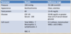
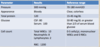
State the pressure, color, total protein, glucose, and cell count for the following:

  1. Bacterial meningitis?
  2. Viral (aseptic meningitis)?
  3. Fungal meningitis?
  4. Multiple sclerosis?
  5. Guillian-Barre’?
  6. SAH?
A

See picture

32
Q

Interpret these results - 1

A

Bacterial Meningitis

33
Q

Interpret these results -2

A

Aseptic (viral) meningitis

34
Q

Interpret these results - 3

A

Fungal meningitis

35
Q

Interpret these results - 4

A

Multiple sclerosis

36
Q

Interpret these results - 5

A

Guillan Barre

37
Q

Interpret these results - 6

A

Subarachnoid hemorrhage

38
Q

What are the two types of nerve conduction studies are there?

A
  1. Nerve conduction velocity
  2. Electromyelography (EMG)
39
Q
  1. Nerve conduction velocity measures what?
  2. What disease processes does it evaluate? 2
A
  1. Measures how well and how fast the peripheral nerves send the signals
  2. Evaluation of
    - entrapment neuropathies
    - Guillian-Barre’
40
Q

What does Electromyelography (EMG) measure?

A

Measures the electrical activity of muscles during rest and contraction

41
Q
  1. Nerve conduction studies test what nerves only?
  2. Can diagnose what kind of disorders? 2
  3. What is its main use? !!!!!!
  4. How would you sort this out?
A
  1. Tests the peripheral nerves only
  2. can diagnose a focal or generalized disorder
  3. Used to differentiate muscle disorders from nerve disorders!!!!

Sorting it out:

  1. Is the nerve signal adequate but the muscle is not responding vs. altered nerve signals and intact muscle response
42
Q

Indications for Nerve conduction studies

3

A
  1. Evaluation of paresthesias of the arms and legs
  2. Evaluation of weakness of the arms and legs
  3. Some disorders that can be diagnosed by nerve conduction studies:
43
Q

What disorders can be diagnosed by nerve conduction studies?

6

A
  1. Carpal tunnel syndrome,
  2. ulnar neuropathy,
  3. spinal disc herniation,
  4. Guillian-Barre’,
  5. peripheral neuropathy
  6. Myasthenia Gravis
44
Q

Classification of nerve fibers:

What are size of the fibers and what do they sense?

  1. A-alpha?
  2. A-delta?
  3. C fibers?
A

1.

Large myelinated fibers

Touch, vibration, position

2.

Small myelinated fibers

Cold and pain sensation

3.

Unmyelinated fibers

Warm and pain

45
Q
  1. Nerve conduction velocity can only study which kind of fibers?
  2. Can appear normal in who?
A
  1. largest A-alpha fibers

Touch, vibration, position

  1. polyneuropathies with primarily small fiber involvement
46
Q

Nerve conduction velocity

2

A

Useful in studying

  1. demyelinating polyneuropathy (Guillain-Barre’ Syndrome) or
  2. focal demyelination (entrapment neuropathies such as carpal tunnel syndrome)
47
Q

Nerve conduction velocity

  1. Normal velocity if myelin is what?
  2. Slow conduction velocity if what?
A
  1. Normal velocity if myelin is intact
  2. Slow conduction velocity if demyelination or destruction of large fibers
48
Q

What does an EMG find damage in? 3

Specific diseases? 3

A

Find diseases that :

  1. damage muscle tissue,
  2. nerves
  3. the junctions between nerve and muscle

Specific diseases:

  1. Herniated disc,
  2. amyotrophic lateral sclerosis (ALS) or
  3. myasthenia gravis
49
Q

Describe the process of EMG?

A

Needle is placed into the muscle (motor unit)

Electrical stimulus delivered and a motor unit action potential is created

50
Q
  1. What nerve conduction tests should we always order together?
  2. Characterizes disorders of the what? 4
  3. These should not be the first tests ordered but should be used to do what?
A
  1. Should always order nerve conduction velocity and electromyelography together
  2. Characterizes disorders of the
    - motor neuron,
    - neuromuscular junction,
    - primary nerve disorder
    - nerve root disorders
  3. These should not be the first tests ordered but should be used to confirm a diagnosis or judge severity of disease
51
Q

Clinical utility of EEG

4

A
  1. To distinguish epileptic seizures from

—Psycogenic spells, syncope, movement disorders, migraine variants

  1. Differentiate organic causes of encephalopathy or delirium from psychiatric causes
  2. Testing for brain death
  3. Determining whether or not to wean anti-epileptic meds
52
Q
  1. Electrodes on the scalp record what?
  2. What is the electrical activity recorded as?
  3. The wave forms are labeled according to their what?
  4. What are the categories of wave forms? 4
A
  1. Electrodes on the scalp record the synaptic activity of the brain
  2. This electrical activity is recorded as a wave form
  3. The wave forms are labeled according to their amplitude

4.

  • Delta 0-4 Hz,
  • Theta 4-8 Hz,
  • Alpha 8-12 Hz,
  • Beta greater than 12 Hz
53
Q
  1. Increased slow wave activity in an awake pt may = what?
  2. With increased slow activity what will we see?
  3. Increased slow wave acitivity is almost always abnormal at what time?
  4. Focal delta activity is usually irregular in configuration and is termed what? What is this indicative of?
A
  1. focal brain lesion
  2. theta and delta waves
  3. Increased slow wave activity when awake is virtually always abnormal
  4. Focal delta activity is usually irregular in configuration and is termed polymorphic delta activity (PDA). PDA is usually indicative of a focal brain lesion.
54
Q
  1. Generalized theta and delta activity is an indication of what?
  2. As a general rule, EEG is a sensitive test for what?
A
  1. encephalopathy
  2. encephalopathy
55
Q
  1. EEG is very useful in the diagnosis of what?
A

seizure disorders

56
Q

EEG techniques that are useful in inducing epileptic activity and increasing test sensitivity are?

5

A
  1. Hyperventilation,
  2. photic stimulation,
  3. sleep,
  4. sleep deprivation, and
  5. drugs,
57
Q

What are the three things that EEGs are used to diagnose?

A

Able to diagnose seizures, encephalopathy, focal brain lesions