17 Acute Kidney Injury Flashcards
1
Q
Definitions
- Acute Kidney Injury (AKI)
- Acute Renal Failure (ARF)
- Azotemia
- Uremia
- Oliguria
- Anuria
A
- Acute Kidney Injury (AKI)
- Loss of renal function, as assessed by GFR, over a period of hours to days
- Hallmark: retention of nitrogenous waste products in the blood
- Often, but not always, accompanied by a reduction in urine volume
- Acute Renal Failure (ARF)
- Older term for AKI
- Now, severe AKI requiring renal replacement therapy
- Azotemia
- Accumulation of nitrogenous waste products (e.g., urea, creatinine) in the blood
- Uremia
- Symptomatic renal failure
- Symptoms: anorexia, nausea, vomiting, muscle cramps, restless legs, sleep disorders, asterixis, mental status changes, seizures, fluid and electrolyte disturbances, anemia, platelet dysfunction and pericarditis
- Oliguria
- Low urine volume
- 24-hour urine volume < 400 to 500 mL
- Volume of 400 to 500 mL = min urine volume required to excrete the obligate daily solute load at a max urinary conc
- Anuria
- Absence of urine
- 24-hour urine volume < 100 mL.

2
Q
Manifestations of AKI
- Primary clinical manifestations
- Conc of urea & creatinine in AKI
- Na & water retention
- K, H, & phosphate excretion
- Excretion of meds or their metabolites
A
- Primary clinical manifestations
- Accumulation of nitrogenous waste products in the blood
- Esp urea (BUN) & creatinine
- Initially: accumulation of substances (azotemia) is asymptomatic
- W/ time: symptoms of renal failure (uremia)
- Accumulation of nitrogenous waste products in the blood
- Conc of urea & creatinine in AKI
- Not necessarily in steady state
- Not possible to estimate GFR from serum concs of these solutes
- Na & water retention
- –> expansion of EC volume & volume overload
- –> edema & pulm vascular congestion or pulm edema
- K, H, & phosphate excretion
- Decreased excretion –> hyperkalemia, metabolic acidosis, & hyperphosphatemia
- Excretion of meds or their metabolites
- Decreased excretion of meds or their metabolites –> accumulation & toxicity

3
Q
Differential diagnosis of azoetmia
- Azotemia
- Most common cause of azotemia
- What causes increased plasma urea conc despite preserved GFR
- What causes increased creatinine conc in the absence of renal failure
A
- Azotemia
- Acute elevations of urea/BUN or creatinine
- Most common cause of azotemia
- AKI
- What causes increased plasma urea conc despite preserved GFR
- Increased urea generation from protein loading
- Protein is metabolized to urea
-
GI bleeding
- Endogenous protein load
-
Catabolic steroids (ex. glucocorticoids)
- Increase protein catabolism
-
Tetracycline antibiotics
- Inhibit protein synth
- Increased urea generation from protein loading
- What causes increased creatinine conc in the absence of renal failure
- Inhibition of tubular secretion of creatinine by meds (ex. cimetidien or trimethoprim)
- Interference w/ colorimetric creatinine assays by meds (ex. cefoxitin, flucytosine, or acetoacetate (ketoic states))
4
Q
Clinical definition & staging of AKI
- Consensus definition of AKI
- Staging of AKI
- Stage 1
- Increase in serum creatinine
- Urine output
- Stage 2
- Increase in serum creatinine
- Urine output
- Stage 3
- Increase in serum creatinine
- Urine output
- Stage 1
- AKI vs. CKD vs. AKD
- AKI
- CKD
- AKD
A
- Consensus definition of AKI
- Increased serum creatinine by > 0.3 mg/dL within 48 hrs
- Increased serum creatinine by > 50% within 7 days
- Urine volume < 0.5 ml/kg per hour for > 6 hrs
- Staging of AKI
- Stage 1
- Increase in serum creatinine: > 0.3 mg/dl or 1.5-2x baseline
- Urine output: < 0.5 ml/kg per hour for 6-12 hrs
- Stage 2
- Increase in serum creatinine: 2-3x baseline
- Urine output: < 0.5 ml/kg per hour for > 12 hrs
- Stage 3
- Increase in serum creatinine: > 3x baseline or > 4 mg/dl or initiation of renal replacement therapy
- Urine output: < 0.3 ml/kg per hour for > 24 hrs or anuria for > 12 hrs
- Stage 1
- AKI vs. CKD vs. AKD
- AKI
- Increase in serum creatinine by > 50% within 7 days
- Increase in serum creatinine by > 0.3 mg/dl within 2 days
- CKD
- GFR < 60 ml/min per 1.73 m2 for < 3 months
- AKD
- AKI
or - GFR < 60 ml/min per 1.73 m2 for < 3 months or decrease in GFR by > 35% or an increase serum creatinine by > 50% for < 3 months
- AKI
- AKI

5
Q
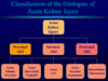
Classification of etiologies of AKI
A
- Prerenal AKI
- Prerenal azotemia
- No structural damage
- Decrease in kidney function is due to hypoperfusion
- Prerenal azotemia
- Intrinsic AKI
- Acute tubular necrosis (ATN)
- Acute interstitial nephritis (AIN)
- Acute glomerulonephritis (AGN)
- Acute vascular syndromes
- Intratubular obstruction
- Postrenal AKI
- Obstructive uropathy
6
Q
Prerenal AKI (prerenal azotemia):
Pathophysiology
- Occurs when…
- Key aspect
- Normal response to decreased perfusion
- Abnormal response to decreased perfusion
- Tubular Na & urea reabsorption in states of decreased renal perfusion w/ maintained GFR & prerenal azotemia
A
- Occurs when…
- Decreased renal perfusion –> decreased GFR
- Exaggeration of the normal physoilogic response to reductions in renal perfusion
- Key aspect
- Absence of histologic changes in the kidney
- Reduction in renal function is entirely hemodynamically mediated
- Normal response to decreased perfusion
- Mediated by AII –> AffA & EffA vasoconstriction
- Release of vasodilatory prostaglandins –> inhibits AII effect on AffA
- Net effect: EffA vasoconstriction w/ minimal/no change in AffA tone
- Maintains glomerular capillayr pressure depsite decreased renal plasma flow
- Preserves GFR at the expense of increased FF
- Abnormal response to decreased perfusion
- Further decreased renal perfusion –> decreased ability to maintain GFR
- Decrease glomerular capillary pressure –> decreased GFR despite elevated FF
- Tubular Na & urea reabsorption in states of decreased renal perfusion w/ maintained GFR & prerenal azotemia
- Increased Na reabsorption –> decreased urine Na
- Increased urea reabsroption –> increased plasma urea : creatinine ratio

7
Q
Prerenal AKI (prerenal azotemia):
Clinical settings in which prerenal AKI occurs
- True volume
- Effective blood volume
- States of renal vasculature
A
- True volume depletion
- Decreased effective blood volume
- CHF
- Cirrhosis
- Nephrotic syndrome
- Sepsis
- States of renal vasoconstriction
- Hypercalcemia
- NSAIDs
- Inhibit AffA dilation in opposition to AII’s effect
- Decrease renal plasma flow & glomerular capillary pressure in states associated w/ increased AII production
- Hepatorenal syndrome
- Intense renal vasoconstriction w/ advanced liver disease
- Resembles other forms of prerenal azotemia but doesn’t reverse w/ IV volume expansion
- Reversal of the liver disease w/ liver transplant (or kidney transplant into a pt w/o liver disease) restores renal function
- Poor prognosis w/o liver transplant

8
Q
Prerenal AKI (prerenal azotemia):
Clinical presentation
- History
- Physical exam
- Typical
- True volume depletion
- CHF
- Liver disease
- Lab findings
- BUN : creatinine
- Urine output
- Urine osmolality
- Renal tubular Na reabsorption
- Urine sediment
A
- History
- Acute volume losses (ex. vomiting, diarrhea, acute blood loss)
- Decompensated CHF, liver disease, or acute infection
- Diuretic use
- Changes in weight
- Thirst
- Orthostatic symptoms (ex. lightheadedness on standing)
- Physical exam
- Typical: HoTN, tachycardia, orthostatic changes, decreased skin turgor, & dry mucous membranes & axillae
- True volume depletion: not distended neck veins, clear lung fields, minimal/no peripheral edema
- CHF: pulm rales, S3 cardiac gallop, peripheral edema
- Liver disease: ascites, peripheral edema
- Lab findings
- BUN : creatinine ratio > 20 : 1
- Passive urea reabsroption from the tubule due to decreased tubular fluid flow rate
- Oliguria < 500 ml / 24 hrs
- May be non-oliguric
- Concentrated urine
- Urine osmolality > 700 mmol/L
- Urine specific gravity > 1.020
- Reflects hemodynamically mediated vasopressin secretion
- Increased renal tubular Na reabsorption
- Urine Na < 20 mmol/L
- Fractoin excretion of Na < 0.01
- Bland urine sediment
- BUN : creatinine ratio > 20 : 1
9
Q
Prerenal AKI (prerenal azotemia):
Fractional excretion of Na (FE<sub>Na</sub>)
- FENa
- Calculation
- FENa in prerenal states vs. ATN in pts w/ oliguria & AKI
- Etiologies of FENa < 0.01
A
- FENa
- Index of renal tubular Na reabsorption
- Differentiates b/n etiologies of AKI
- Calculation
- FENa = excreted Na / filtered Na
- Excreted Na = urine Na (UNa) * urine volume (V)
- Filtered Na = plasma Na (PNa) * GFR
- FENa = (UNa * V) / (PNa * GFR)
- GFR = [urine creatinine (UCr) * V] / plasma creatinine (PCr)
- FENa = (UNa / PNa) / (UCr / PCr)
- FENa = excreted Na / filtered Na
- FENa in prerenal states vs. ATN in pts w/ oliguria & AKI
- Prerenal FENa < 0.01
- ATN FENa > 0.02
- Etiologies of FENa < 0.01
- Normal renal function
- Prerenal azotemia
- Hepatorenal syndrome
- Early obstructive uropathy
- Contrast nephropathy
- Rhabdomyolysis
- Acute glomerulonephritis
10
Q
Prerenal AKI (prerenal azotemia):
Treatment
- Primary treatment
- Discontinue…
- In pts w/ underlying heart disease
A
- Primary treatment
- Correction of volume deficits via administration of crystalloid soln’s
- Discontinue…
- Diuretics
- Meds that alter intrarenal hemodynamics (NSAIDs, ACE-Is, ARBs)
- In pts w/ underlying heart disease
- Optimize cardiac function w/ inotropic support &/or vasodilators
11
Q
Postrenal AKI (obstructive uropathy):
Definition
- Results from…
- Hallmark
- Upper vs. lower tract obstruction
- Unilateral upper tract obstruction
- For upper tract obstruction to cause AKI…
- Complete vs. partial obstruction
A
- Results from…
- Partial or complete obstruction of hte urinary tract b/n the renal pelvis & urethral meatus
- Hallmark
- Hydronephrosis (dilation of the renal collecting system)
- Upper vs. lower tract obstruction
- Upper: above the urinary bladder (ex. ureters, renal pelvis)
- Lower: at the bladder outlet or urethra
- Unilateral upper tract obstruction
- Hydronephrosis will be present
- Serum creatinine ill be normal or minimally elevated due to continued function of hte contralateral kidney
- For upper tract obstruction to cause AKI…
- Obstruction must be bilateral
- Unilateral obstruction will only cause AKI if the contralateral kidney is absent or nonfunction
- Complete vs. partial obstruction
- Complete –> anuria
- Partial –> urine flow that’s normal, decreased (oliguria), increased (polyuria), or fluctuating b/n oliguria & polyuria

12
Q
Postrenal AKI (obstructive uropathy):
Pathophysiology
- Initially
- Later
A
- Initially
- Obstruction in the renal collecting system
- –> increased intratubular pressure in the nephron
- –> increased hydrostatic pressure in bowman’s space
- –> increased renal plasma flow –> increased glomerular capillary pressure
- –> decreased gradient b/n pressures in the glomerular capillary & bowman’s space –> decreased GFR
- –> initial increased renal plasma flow decreases –> decreased glomerular capillayr pressure –> decreased GFR
- Later
- Intratubular pressure returns to normal
- –> decreased renal plasma flow –> decreased glomerular capillary pressure & GFR
13
Q
Postrenal AKI (obstructive uropathy):
Etiologies
- Upper tract obstruction
- Intrinsic obstruction
- Extrinsic obstruction
- Lower tract obstruction
A
- Upper tract obstruction
- Intrinsic obstruction
- Nephrolithiasis
- Papillary necrosis
- Blood clot
- Transitional cell carcinoma
- Extrinsic obstruction
- Retroperitoneal or pelvic malignancy
- Retroperitoneal adenopathy
- Retroperitoneal fibrosis
- Endometriosis
- Abdominal aortic aneurysm
- Intrinsic obstruction
- Lower tract obstruction
- Benign prostatic hypertrophy
- Prostate cancer
- Transitional cell carcinoma
- Urethral stricture
- Bladder stones
- Blood clots
- Neurogenic bladder
14
Q
Postrenal AKI (obstructive uropathy):
Clinical presentation
- History
- Frequently
- May present w/
- Bladder outlet obstruction
- Upper tract disease
- Either upper or lower tract disease
- Important to elicit
- Physical exam
- Lab findings
- BUN : creatinine
- Urine sediment
- Urine chemistries
A
- Hisotry
- Frequently: no complaints
- May present w/: anuria, polyuria, widely fluctuating urine volume
- Bladder outlet obstruction: urinary frequency, urgency, intermittency, hesitancy, nocturia, incomplete voiding
- Upper tract disease: flank pain (ureteral colic)
- Either upper or lower tract disease: hematuria
- Important to elicit: pelvic malignancy, radiation therapy, prostate disease
- Physical exam
- Distended bladder palpable as a suprapubic mass
- Prostatic enlargmeent
- Pelvic masses
- Adenopathy
- Lab findings
- BUN : creatinine > 20 : 1 (variable)
- Urine sediment
- Often unremarkable
- Microscopic hematuria (w/o RBC casts) may be present
- Crystaluria may be seen w/ nephrolithiasis
- Urine chemistries (variable & non-diagnostic)
15
Q
Postrenal AKI (obstructive uropathy):
Diagnostic studies
- Post-void residual bladder volume
- Radiologic studies
- Renal ultrasound
- CT scan
- Nuclear medicine excretory renogram
- Retrograde pyelography
- Antegrade nephrostogram
A
- Post-void residual bladder volume (lower tract obstruction)
- Measured by placing a bladder catheter or using an ultrasound device to measure residual bladder volume after having pt void & completely empty bladder
- Residual bladder volume > 100 ml –> voiding dysfunction
- Radiologic studies (upper tract obstruction)
- Renal ultrasound: initial imaging study
- CT scan: best initial study for kidney stones
- Nuclear medicine excretory renogram: functional test, used to define if urinary tract dilatoin is due to obstruction
- Retrograde pyelography: invasive, may be accompanied by placement of ureteral stent for treatment
- Antegrade nephrostogram: invasive, usually accompanied by placement of percutaneous nephrostomy for treatment

17
Q
Intrinsic AKI:
Acute tubular necrosis:
Etiologies
- General
- Ischemic ATN
- Nephrotoxic ATN
- Exogenous toxins
- Endogenous toxins
A
- General
- Most common form of intrinsic AKI (85% of cases)
- Etiology is frequently multifactorial
- Ischemic ATN
- Prolonged prerenal azotemia
- Hypotension
- Hypovolemic shock
- Cardiopulmonary arrest
- Cardiopulmonary bypass
- Sepsis
- Nephrotoxic ATN
- Exogenous toxins (Drug-induced)
- Radiocontrast agents
- Aminoglycoside antibiotics
- Amphotericin B
- Cisplatinum
- Acetaminophen
- Endogenous toxins (Pigment nephropathy)
- Hemoglobin (intravascular hemolysis)
- Myoglobin (rhabdomyolysis)
- Exogenous toxins (Drug-induced)

18
Q
Intrinsic AKI:
Acute tubular necrosis:
Pathophysiology
- Animal vs. human pathology
- At the cellular level, nephrotoxic or ischemic injury –>
- Ultimately…
- Loss of GFR is due to…
- Recovery of renal function
A
- Animal vs. human pathology
- Animals: widespread cell necrosis
- Humans: patchy cell necrosis that doesn’t necessarily correlate w/ severity
- At the cellular level, nephrotoxic or ischemic injury –>
- Loss of normal tubular epithelial cell morphology
- –> loss of normal apical brush border
- –> loss of cellular polarity (differentiation b/n apical & basolateral membrane domains)
- Migration of transport proteins usually restricted to the basolateral membrane (ex. Na/K ATPase) into the apical membrane or vice versa (ex. Na/H exchanger)
- Loss of epithelial cell polarity disrupts tubular transports
- Also renal vasoconstriction & endothelial cell injury
- Ultimately…
- Epithelial cel ldeath occurs due to necrosis & apoptosis
- Viable & dead cells slough into the tubular lumen –> areas of denuded tubular BM
- Sloughed cells & debris form epithelial cell casts which may obstruct the tubular lumen
- Loss of GFR is due to…
- Renal vasoconstriction
- Tubular obstruction from sloughed debris
- Back-leak of glomerular ultrafiltrate across teh denuded tubular BM
- Recovery of renal function
- Remaining viable epithelial cells dedifferentiate, proliferate, & spread across the denuded BM
- Re-differentiation & re-establishment of cell polarity

19
Q
Intrinsic AKI:
Acute tubular necrosis:
4 phases of endothelial injury & activation of inflammatory pathways
A
- Initiation
- Acute ischemic event
- Extension
- Initiating event has resolved
- Endothelial injury, vasoconstriciton, & activation of inflammatory mediators –> continued tubular injury
- Maintenance
- Continued ifnlammation
- Resolution of endothelial injury
- Initiation of tubular repair
- Migration of epitehlial cells over denuded BM
- Recovery
- Proliferatoin & re-differentiatoin of epithelial cells
- Recovery of GFR toward normal

20
Q
Intrinsic AKI:
Acute tubular necrosis:
Clinical presentation
- History
- Physical exam
- Lab adata
- BUN : creatinine
- Urine volume
- Urine osmolality
- Renal Na
- Urine sediment
A
- History
- Acute illnesses
- Meds
- Exposure to other nephrotoxins
- Episodes of HoTN
- Physical exam
- Hemodynamic status
- Volume status
- Features of associated illness
- Lab data
- BUN : creatinine < 10 : 1
- Urine volume
- Oliguric or non-oliguric
- Isothenuric urine
- Urien osmolality ≈ 300 mmol/L
- Specific gravity ≈ 1.010
- Renal Na wasting
- Urine Na > 40 mmol/L
- FENa > 0.02
- Urine sediment
- Tubular epithelial cells & epithelial cell casts
- Granular casts (“muddy” brown casts)

22
Q
Intrinsic AKI:
Acute interstitial nephritis:
General & pathology
- Aka
- General
- Classic presentation
- Pathology
A
- Aka
- Allergic interstitial nephritis
- General
- Immune mediated form of AKI
- Lymphocytic infiltration of interstitium
- Accounts for 5-10% of intrinsic AKI cases
- Classic presentation (30% of cases)
- AKI w/ triad of fever, rash, & eosinophilia
- Pathology
- Lymphocytic infiltrate
- Frequently accompanied by eosinophils

24
Q
Intrinsic AKI:
Acute interstitial nephritis:
Clinical presentation
- History
- Physical exam
- Lab findings
- Urine findings
A
- History
- Preceding illness or drug exposure
- Physcial exam
- Fever
- Rash
- Lab findings
- Eosinophilia
- Urine findings
- Non-nephrotic proteinuria
- Hematuria
- Pyuria
- WBC casts
- Eosinophiluria
- Non-specific finding
- Don’t need to see them to make the diagnosis
- Absence doesn’t rule out the diagnosis
- Positive predictive value of ~ 50%
- Negative predictive value of > 90%
- Non-specific finding

26
Q
Intrinsic AKI:
Acute glomerulonephritis
- General
- Presentation
- Exact diagnosis requires…
- Etiologies
A
- General
- Accounts for 5-10% of intrinsic AKI cases
- Presentation
- Nephritic urine sediment
- Hematuria
- RBC casts
- Exact diagnosis requires…
- Kidney biopsy
- Etiologies
- Poststreptococcal glomerulonephritis
- Postinfectious glomerulonephritis
- Endocarditis-associated glomerulonephritis
- Systemic vasculitis
- Thrombotic microangiopathy
- Hemolytic-uremic syndrome (HUS)
- Thrombotic thrombocytopenic purpura (TTP)
- Rapidly progressive glomerulonephritis (RPGN)
27
Q
Intrinsic AKI:
Acute vascular syndromes
- General
- Examples –> renal infarction
- Irreversible AKI
- Atheroembolic disease
- Atheroembolism
- Cutaneous findings
- Initial shower of emboli
- Clinical course of kidney disease
- Pathognomonic finding
- Atheroembolic disease frequently occurs after…
A
- General
- Uncommon forms of AKI
- Examples –> renal infarction
- Renal artery thromboembolism
- Renal artery dissection
- Renal vein thrombosis
- Irreversible AKI
- Bilateral infarction
- Infarction affects a single functioning kidney
- Atheroembolic disease
- Aka cholesterol embolism
- Results from rupture of atheromatous plaques in the aorta w/ embolization of atheromatous material into distal arterioles
- May affect many organ systems (ex. GI tract, muscle & skin, kidney)
- Cutaneous findings
- Livedo reticularis
- Micro-infarcts of the digits (blue-toe syndrome)
- Initial shower of emboli
- Usually doesn’t result in vascular occlusions
- Triggers a secondary inflammatory response –> fibrosis
- Clinical course of kidney disease
- Frequently sub-acute (days to weeks) rather than acute
- Pathognomonic finding
- Biconcave, needle-shaped clefts on histologic exam of blood vessels
- Result from the dissolutoin of the cholesterol crystals int eh emboli during tissue fixation
- Atheroembolic disease frequently occurs after…
- Antiographic procedures
- Frequently needs to be differentiated fromteh more common radiocontrast-inducted ATN

28
Q
Intrinsic AKI:
Intratubular obstruction
- General
- May result from…
A
- General
- Uncommon form of AKI
- May result from…
- Crystaline material
- Uric acid in tumor lysis syndrome
- Ca oxylate following ethylene glycol ingestion or drug crystals (ex. acyclovir)
- Meds (ex. acyclovir)
- Proteinaceous material
- Light chain cast nephropathy in multiple myeloma (ex. Bence-Jones protein deposition)
- Crystaline material
