Abdominal Anatomy Flashcards
Four abdominal fascial layers
- Superficial fascia (subcutaneous tissue), which lies underneath the skin and contains various amount of fat.
- Investing fascia, which covers the external aspect of the abdominal muscles.
- Endo-abdominal fascia or transversalis fascia, which covers the internal aspect of the abdominal muscles.
- Extraperitoneal fat, which separates the endo-abdominal fascia and the peritoneal layer that covers the abdominal wall (parietal peritoneum).

Muscles of the anterolateral abdominal wall

Flat abdominal wall muscles
- External abdominal oblique is the superficial muscle.
- Internal abdominal oblique is the intermediate muscle.
- Transversus abdominis is the innermost muscle.

Vertical abdominal wall muscles
- Rectus abdominis is a long strap-like muscle that is mostly surrounded by the rectus sheath.
- Pyramidalis is a short triangular muscle that lies inside the rectus sheath.

Contents of the abdominal rectus sheath
- Rectus abdominis muscles
- Pyramidalis muscles
- Superior and inferior epigastric arteries and veins, lymphatic vessels
- Thoracoabdominal and subcostal nerves

Anterior abdominal arteries

Anterior abdominal veins

Nervous supply of the anterior abdominal wall
Branches of the T7-T12 and L1 spinal nerves supply the anterolateral abdominal wall.
Hesselbach’s Triangle
Common site of hernia.
Bordered medially by the rectus abdominus muscles, superiorly by the inferior epigastric vein and artery, and inferiorly by the external inguinal ring and inguinal ligament.

Inguinal canal
The inguinal canal is an inferomedially directed oblique passage that is formed during fetal development when the gonads are relocated from the dorsal abdominal wall to their final destination.
Borders of the inguinal canal
- The anterior wall is formed by the aponeurosis of the external abdominal oblique muscle.
- The posterior wall is formed by the transversalis fascia, which is reinforced by the aponeuroses of the internal abdominal oblique and transversus abdominis muscles.
- The roof is formed by the transversalis fascia and the internal abdominal oblique and transversus abdominis muscles.
- The floor is formed by ligaments (inguinal and lacunar).

Inguinal canal openings
- The deep (internal) ring is the entrance of the inguinal canal. It is formed by the invagination of the transversalis fascia.
- The superficial (external) ring is the exit of the inguinal canal. It is a narrow opening in the aponeurosis of the external abdominal oblique muscle.

Inguinal canal contents
In male: spermatic cord, genital branch of the genitofemoral nerve and ilioinguinal nerve
In female: round ligament of the uterus, genital branch of the genitofemoral nerve and ilioinguinal nerve
Pain on the parietal peritoneum is generally ____ localized.
Pain on the parietal peritoneum is generally well-localized.
Unlike the underlying tissues, the peritoneum is innervated by the same somatic nerve supply as the abdominal wall, and so it produces a much more location-specific feeling that is typical of somatic sensation as opposed to the dull, vaguely-localized pain associated with visceral sensation.
Pain on the visceral peritoneum is generally ____ localized.
Pain on the visceral peritoneum is generally vaguely-localized.
The visceral peritoneum, unlike the parietal peritoneum, is innervated by visceral sensory nerve fibers, and so it produces a dull and unspecifically localized pain.
Visceral and parietal peritoneum

Intraperitoneal organs
liver, stomach, spleen
Early embryonic dorsal and ventral mesogastrium of the foregut
As the embryo develops, the dorsal and ventral mesogastria curve, the liver develops in the ventral mesogastrium, and the spleen develops in the dorsal mesogastrium.
The liver grows rapidly and eventually induces a separation of the peritoneal sac against the right anterior abdominal wall. This produces the lesser peritoneal sac, which in turn gives rise to the lesser omentum.

Foregut structures
The foregut derivatives are the esophagus, stomach, pancreas, duodenum up to papilla of Vater (greater duodenal papilla), liver and biliary ducts.
Midgut structures
The midgut derivatives are the small intestines (starting with duodenum after papilla of Vater), cecum, appendix, ascending colon and majority of the transverse colon.
Hindgut structures
The hindgut derivatives are the distal part of the transverse colon, descending colon, sigmoid colon and rectum.
Embryological rotation of the midgut
Gives the characteristic weaving of the gut we see in adults, where the transverse colon (part of the midgut) extends anterior to the duodenum.
At week 10 the midgut rotates 270 degree around the axis of its supporting blood vessel (superior mesenteric artery). As the parts of the intestines reach their final position, their peritoneal coverage changes (some part will be shorter, others will disappear).
Peritoneal development
At 2-3 weeks of development the embryonic body cavity is lined by mesoderm. At 1 month, the primordial abdominal cavity is lined with parietal peritoneum (derived from mesoderm) that forms a closed sac.
The lumen of this sac is the peritoneal cavity. As the abdominal organs develop and grow they protrude to varying extent into the peritoneal sac. The part of the peritoneum that covers the organs is the visceral peritoneum.
The abdominal organs are ___ to the abdominal wall
The abdominal organs are connected to the abdominal wall
through varying length of peritoneal formations (such as the mesentery). The longer the peritoneal support the more flexible is the organ. As organs protrude into the peritoneal sac they remain connected to their neurovascular supply, so these structures lie between the layers of the peritoneum.

fusion fascia
Fascia that forms as a result of the compression of visceral and parietal peritoneum.
Generation of fusion fascia is important to peritoneal development, particularly the fusion fascia of the liver and anterio-lateral abdominal wall and of the descending colon and posterio-lateral abdominal wall.
Organs that have only partial covering of peritoneum are “secondarily retroperitoneal”.
Secondarily retroperitoneal organs
Secondarily retroperitoneal organs are most of the duodenum, ascending, descending colon and the pancreas.
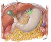
Lesser and greater omentum on the stomach.

Falciform ligament and ligamentum teres
Descend from the abdominal ceiling over the liver and then connect to the umbilicus. The ligamentum teres represents the remains of the umbilical vein.

Mesentery
The mesentery is a double layer of peritoneum that is formed when the organs invaginate the peritoneum during development. Thereafter, the mesentery provides the neurovascular communication between the organ and the abdominal wall. The mesentery also connects the intraperitoneal organ to the body wall (usually to the posterior abdominal wall).

Peritoneal fold
A peritoneal fold is a reflection of the peritoneum that is created when a structure (e.g. blood vessel or duct) lifts the peritoneum from the abdominal wall. Some peritoneal folds are called peritoneal ligaments when they connect an organ to the abdominal wall or to another organ.

The falciform ligament
connects the liver to the anterior abdominal wall.

The hepatogastric ligament
connects the liver and the stomach; it is part of the lesser omentum.

The hepatoduodenal ligament
connects the liver and the duodenum; it is part of the lesser omentum.

gastrophrenic ligament
connects the stomach and the inferior surface of the diaphragm. Part of the greater omentum

gastrosplenic ligament
connects the stomach and the spleen. Part of the greater omentum.

The gastrocolic ligament
The gastrocolic ligament connects the stomach to the transverse colon. Part of the greater omentum

Bare area of the liver
(Picture shown is a view form the posterior)
Area uncovered by the peritoneum that provides a space for neurovascular supply.

Paracolic gutters

Rectouterine pouch

Rectovesical pouch

Greater and Lesser peritoneal sacs

Cardia of the stomach
Surrounds the cardiac orifice, which is the trumpet-shaped opening of the esophagus into the stomach.
Fundus

Body of the stomach

Pylorus of stomach

Duodenum position

Jejunum and ileum positions

Parts of the large intestine

Tactile features of the large intestinal tract

Parts of GI tract on axial radiograph

Liver (front)

Liver (back)

View inside the liver
Note that the lobes are divided into 8 segments.

Ligaments anchoring the liver

Hepatorenal recess

Liver bile ducts

Sections of the pancreas

Location of the pancreas

Visceral Surface of the Spleen

Splenic ligaments

Splenorenal recess
