CNS 1 Flashcards Preview

Physiology > CNS 1 > Flashcards

Flashcards in CNS 1 Deck (60)
Loading flashcards...
1
Q

Parts of nervous system

A
2
Q

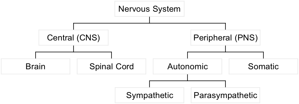
Central nervous system

A

Brain and spinal cord

3
Q

peripheral nervous system

A

Autonomic and somatic

4
Q

sympathetic nervous system

A
  • part of autonomic nervous system
  • fight or flight
5
Q

parasympathetic

A

rest and digest

6
Q

somatic

A
  • part of peripheral nervous system
  • voluntary control of skeletal muscles
7
Q

white matter

A

high density of myelin covering axon pathways (and very few neurons)

8
Q

gray matter

A

high density of neurons and dendrites (axons also present)

9
Q

nucleus

A

cluster of neurons within the CNS

10
Q

ganglion

A

cluster of neurons outside of the CNS

11
Q

cortex

A

dense layers of neurons

12
Q

tract

A

axons within the CNS traveling as a group/usually named based on region of origin and termination

i.e. spinocerebellar tract

13
Q

pathway

A

similar to tract however it relates more to synaptically connected neurons performing a function

14
Q

spinal cord function

A
  • associated nerves: Dorsal and ventral roots
  • primary function:
    • sensory input
    • reflex circuits
    • somatic and autonomin motor output
15
Q

dorsal root

A
  • posterior
  • sensory
  • afferent (towards the brain)
  • joins with the ventral root to form the spinal nerve
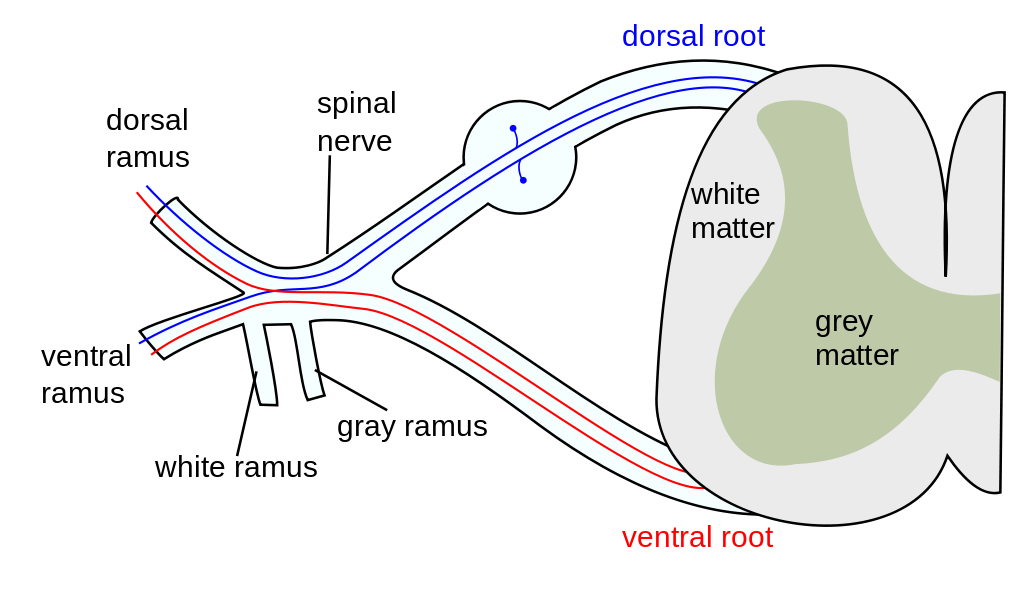
16
Q

ventral root

A
  • anterior
  • efferent (exiting the brain)
  • motor
  • joins with dorsal root to form spinal nerve
17
Q

brainstem

A
  • medulla, pons, and midbrain
  • 12 associated cranial nerves
18
Q

reticular formation

A
  • part of brainstem
  • receives a summary of much of the information that enters the spinal cord and brain stem.
    • filters information and excludes irrelevent stimuli
  • regulates arousal
19
Q

medulla

A
  • Associated nerves:
    • cranial VIII-XII
  • functions:
    • subconscious CV and respiratory control
    • early relay nuclei in auditory, balance/equilibrium, taste
    • head and neck control
    • brainstem reflexes
    • sets baseline tone for blood vessels
20
Q

Pons

A
  • Associated nerves:
    • Cranial nerves V-VIII
  • functions:
    • respiratory control
    • urinary control
    • motor control of the eye
    • sensation and motor control of the face
    • ventral:
      • pontine nuclei relay movement and sensation info from cortex to cerebellum
    • dorsal:
      • taste and sleep
21
Q

pontine nuclei

A
  • ventral (motor) part of pons- relays movement and sensation information from cortex to cerebellum
22
Q

Midbrain

A
  • Associated nerves:
    • Cranial nerves III-IV
  • functions:
    • acoustic relay and mapping (processing hearing)
    • eye movement, lens, and pupil reflex
    • pain modulation
    • contains nuclein and relay pathways critical for motor coordination
      • i.e. substantia nigra
23
Q

Cerebellum

A
  • Associated nerves:
    • cranial nerve VIII
  • function:
    • coordination and equilibrium
      • helps make smooth, coordinated muscles
    • motor learning
    • sensory association/language
  • **found to not be necessary for life
24
Q

thalamus

A
  • associated nerves:
    • cranial nerve II
  • functions:
    • sensory and motor relay/coordination btw cerebral hemispheres and lower CNS regions
    • sensory modulation and gating
    • regulation of cortical activation (attention and consciousness)
      • without thalamus engaged, we can’t be conscious
    • visual input
25
Q

hypothalamus

A
  • primary functions:
    • autonomic/endocrine control
      • controls all pituitary secretions
    • superchiasmatic nucleus
      • circadian rhythms (hormones for day/night)
    • motivated behavior
      • reward centers
26
Q

basal ganglia

A
  • functions:
    • receives fibers from substantia nigra
    • shaper patterns of thalamocortical motor inhibition
      • keeps movements smooth
    • controls fine motor
27
Q

amygdala

A
  • function
    • social behavior and expression of emotion
28
Q

hippocampus

A
  • primary functions:
    • memory
29
Q

cerebral cortex

A
  • associated nerves:
    • CN I
  • dependent on lower brain for “wakefulness”
  • functions:
    • fine tune lower brain functions
    • sensory perception
    • cognition
    • learning
    • large “memory storehouse”
    • motor planning and voluntary movement
    • language
    • planning, personality
30
Q

Cerebral cortex

Frontal lobe

A
  • planning and carrying out motor behavior
  • speech generation (Broca’s area)
  • personality and emotional behavior
31
Q

Cerebral Cortex

Parietal lobe

A
  • sensory perception and processing
  • interconnectal with frontal lobe
    • to connect what is being sensed with motor function
  • Werkicke’s area- understanding speech
32
Q

Occipital lobe

Cerebral cortex

A
  • visual perception and processing
  • controls eye movements, pupil constriction
33
Q

temporal lobe

cerebral cortex

A
  • sound and balance
  • difficult visual processing (i.e. facial recognition)
  • emotional behavior
  • ANS regulation
  • learning and memory
34
Q

circle of willis

A

connection of vessels that supply blood to brain

**lots of collateralization

35
Q

Artery of Adamkiewicz

A
  • supplies blood to lower two thirds of spinal cord
  • comes off of main artery at a different spot on every person with wide variety
    • can be a problem when clamping arter and losing blood flow to most of spine.
36
Q

spinal cord blood supply

A
  • anterior spinal artery
    • only one, no backup
    • supplies anterior 2/3
    • during spinal surgeries we check for motor to check integrity of anterior spinal artery to make sure cord is still being perfused.
  • posterior spinal arteries
    • there are two
    • supply posterior one third
37
Q

cerebral blood volume

A
  • CBV = 3-5ml/100g brain tissue
  • cerebral flow influences total cerebral volume
  • consider arterial flow and venous drainage
    • obstruction to outflow can increase CBV
      • obstruction can be position, vent with high PIP
38
Q

intracranial pressure

A
  • normal 8-12 mmHg
  • measured in lateral ventricles
39
Q

cranial vault fixed volume

A
  • brain (cellular and ICF) = 80%
    • fixed, can’t manipulate the brain
  • Blood (arterial and venous) = 12%
    • we can manipulate this
  • CSF = 8%
    • can also manipulate (ventric, lumbar drains)
    • don’t do this in OR very often
40
Q

ICP curve

A
  • once compensation abilities is maxed out, ICP will increase significantly
    • high ICP decreases CPP
    • herniation
41
Q

Intracranial Elastance (compliance)

A
  • determined by the change in ICP after a change in intracranial volume
  • compensatory mechanisms are:
    • displacement of CSF from cranial to spinal compartment
    • Increased CSF absorption
    • Decreased CSF production
    • Decreased CBV (mostly venous)
42
Q

CPP

A
  • CPP = MAP-ICP (or CVP, whichever is greater)
  • normal is 80-100
43
Q

Cerebral Blood flow

A
  • Normal Adult 50ml/100g/min = 750 ml/min
    • grey matter: 80 ml/100g
    • white matter: 20 ml/100g
  • flow is closely linked with metabolism
    • increased activity, increased metabolism, increased flow
44
Q

Important factors impacting CBF during anesthesia

A
  1. anesthetic agents- cause dilation but decrease activity
  2. level of arousal
  3. metabolic by-products- Ca, K, CO2, NO, lactate; important in regulating vessels
  4. blood viscosity–thicker wont flow as well
  5. temperature- change electrophysiology req
  6. Concentration of CO2 and H ions
  7. O2
45
Q

effect of CO2 on CBF

A
  • increased CO2, increased CBF
  • Most powerful tool we have to quickly change ICP in OR
    • only lasts for about 6 hours before HCO3 normalized pH
    • useful with volatile anesthetics
  • CO2 + H2O = carbonic acid
  • carbonic acid disassociates into H+
  • for each 1 mmHg change in PaCO2:
    • CBF changes 1-2 ml/100g/min
    • CBV changes 0.5 ml/100g brain tissue
46
Q

Brain metabolism

A
  • only 2% of total body mass, 15-20% of total body metabolism and cardiac output
47
Q

Cerebral metabolic rate (CMRO2)

A

3-3.8 ml/100g/min = 50 ml/min of O2

48
Q

Pediatric Cerebral Metabolic Rate (CMRO2)

A

5.2 ml/100g/min

49
Q

brain glucose consumption

A

5.5 mg/100g/min

50
Q

Brain tissue O2 use

A

3.5 ml O2/100g brain tissue

**stays within narrow range except in case of intense brain activity

51
Q

Autoregulation of CBF

A

Between MAP of 70-150, no change to blood flow occurs.

52
Q

Outside of Autoregulation of CBF concens

A
  • outside of auto-regulation for CBF range
  • below normal range:
    • worry about ischemia
  • above normal range:
    • worry about bleeds
53
Q

Cerebral blood flow graph

A
  • as O2 concentration drops below about 60 mmhg, CBF increases
  • As MAP increases, CBF increases (outside of autoregulation)
  • As CO2 increases, CBF increases
    • this is best tool to control CBF
54
Q

Temperature and CBF

A
  • CBF changes 5-7% per 1 degree C of temp change
    • hypothermia decreases CBF and CMRO2
    • hyperthermia increases CBF and CMRO2

**evidence does NOT support the use of hypothermia <35 degrees C without CP bypass

55
Q

CMRO2

A

cerebral metabolic rate

56
Q

Viscosity and CBF

A
  • decrease in HCT will increase CBF but decrease O2 carrying capacity of the blood
57
Q

Cerebral spinal fluid (CSF) information

A
  • CNS “lymphatic system” and protection from mechanical force
  • Formed from choroid plexuses at 0.35 ml/min
    • complete turnover 3-4 times per day
    • about 500 ml per day
  • reabsorbed by arachnoid villi
    • these function like one way valves; with increased CVP, decreased ability to absorb CSF
58
Q

CSF electrolytes

A
  • Na 148
  • K 2.9
  • Cl 120-130
  • Glucose 50-75
  • protein 15-45
  • pH 7.3
59
Q

flow of CSF

A
  1. Fluid from lateral ventricles passes through intraventricular foramina to the third ventricle
  2. Additional fluid is added and then it flows downward along the Aqueduct of Sylvius into 4th ventricle
  3. More fluid added and then it passes through three small openings ( two lateral foramna of Luschka and a midline foramen of Magendie) and enters the Cisterna Magna (a large fluid space that lied behind the medulla and is part of the subarachnoid space)
60
Q

Blood brain barrier

A
  • exists in tissue capillary membranes in all parts of the blood except hypothalamus, pituitary, and area postrema
  • Large molecules and charged ions can’t get through
  • tight junctions between CNS capillary and endothelial cells
    • wholes are much smaller than in other areas
  • astrocytes restrict movement by taking up potassium ions