What modalities are used in neuroimaging?
What are their benefits/ uses/ disadvantages?
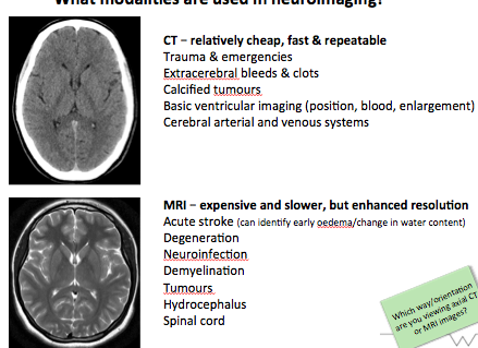
- CT scan:
- Cheap, quick and repeatable
- Emergency and trauma
- extracerebral bleeds and clots (extracerebral meaning any bleed outside cerebrum –> subarachnoid/ subdural/ extradural)
- Calcified tumours
- basic ventricular imaging –> size/ position/ blood
- cerebral arterial and venous system
- MRI:
- Slower, more expensive but high resolution imaging
- Acute stroke –> oedema
- Degeneration and demyelination
- Tumours
- Neuroinfection
- Hydrocephalus
- Spinal cord
What is an MRI?
What are the different strengths of MRI that can be used and what are their advantages/ disadvantages?
- During MRI a strong magnet us used to create radio waves that are directed towards the patient. Radio waves are used to send signals to the body and then receive them back, these returning signals are converted into an image by a computer attached to the MRI
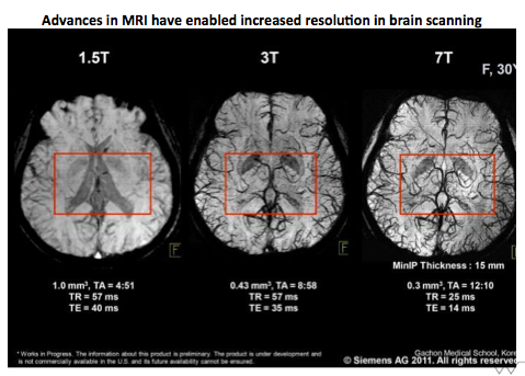
- MRI scanners can have different magnetic field strengths which is measured in Teslas “T”.
- The higher the teslas the higher the resolution image and more detail you will pick up, however there is also an increased chance of artifacts.
- 1.5 T useful for chest and abdominal scans
- 3 T is ideal for imaging small bones, breast MRI, musculoskeletal MRI, neurological and vascular tissues where minute detail is crucial to diagnosis. Most hospitals have 3T MRI.
How might the dural layers be used as landmarks in neuroimaging?
What sinuses are located with the dural reflections?
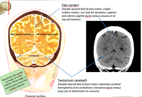
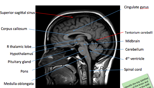
- Know at several points dural reflections occur, separating the different regions of the brain:
- Falx cerebri between R and L hemispheres
- Falx cerebelli between R and L cerebellar hemispheres
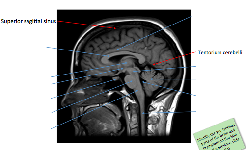
- Tentorium cerebelli overlying the cerebellum and separating occipital lobe.
- This is a useful landmark as Falx cerebri should always be in the midline, if it is shifted this is indicative of a space occupying lesion
- Useful as if the tentorium cerebelli can be seen you know you are far back in the image.
- Sinuses –> falx cerebri associated with superior sagittal sinus and inferior sagittal sinus
- Sinuses –> transverse sinuses associated with tentorium cerebelli
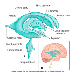
Label the ventricular system
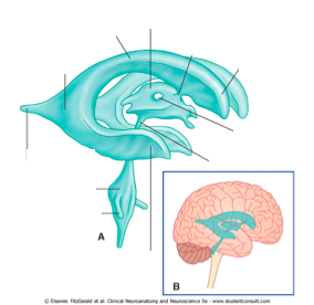
Lateral ventricles split into an anterior horn, body, atrium, occipital horn and temporal horn.
Lateral ventricles drain into the 3rd ventricle via the interventricular foramen
CSF flows from the 3rd ventricle into the 4th ventricle via the cerebral aqueduct.
How can the ventricular system be used to landmark in neuroimaging?
How do they appear in an MRI?
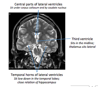
- Ventricles filled with CSF therefore appear white in MRI scan that is weighted T2 or above. Note that different magnet strengths will make images appear differently, therefore on a T1 fluid will not appear as bright.
- Useful landmarks as the central regions of the lateral ventricles are bounded by the caudate nucleus laterally and the corpus callosum superiorly.
- The lateral ventricles lead into the 3rd ventricle which is bound laterally by the two thalamic lobes.
- The inferior portions of the lateral ventricles (i.e the temporal horns) are useful landmarks for the hippocampus.
What is a CT scan?
What modality does it use?
How do images appear?
CT = Computed tomography or CAT scan = Computed Axial Tomography –> same thing.
CT scans based on X-rays, takes sequential pictures of the body as it rotates around. Computed takes data from the single images and combines it with known angle and position to recreate 3D image of the body.
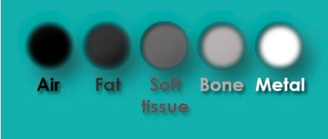
As CT based on XRAY therfore:
bones = white
air = black.
Label the image
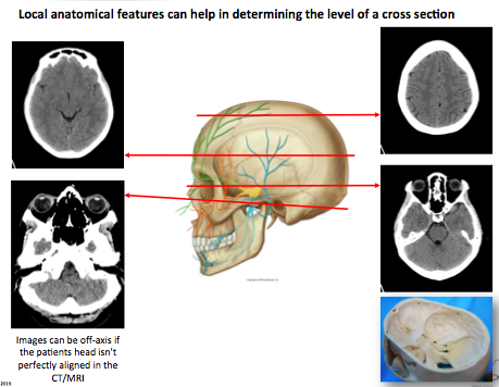
What can help determine the level of a cross section in neuroimaging?
- You need to look for local anatomical features which will help orientate what level you are at:
- Top L image –> frontal sinuses, therefore know you’re just above the eyebrows
- Middle R image –> Passing through the orbits
- Bottom L image–> External auditory meatus and cerebellum visible therefore know youre lower level.
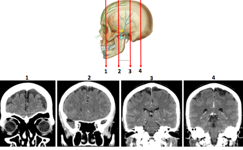
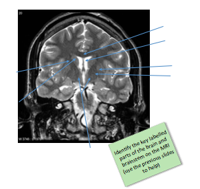
What key anatomical features help distinguish the plane of the section?

- Image 1: orbits, frontal sinus, ethmoid cells and maxillary sinus
- Image 2: nasal septum, nasal cavities, mandible
- Image 3: lateral ventricles, anterior horns and 3rd ventricle
- Image 4: lateral ventricles, brainstem dropping via foramen magnum
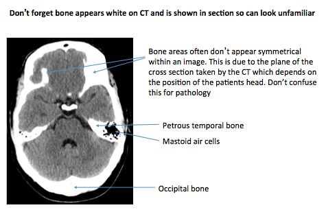
label the image
What type of image is this?
Why is there asymmetry?
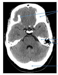
The image is a CT scan, top two arrows point towards bone as bone appears white in CT scans.
The asymmetry is due to the plane of the cross secton and isn’t actually pathology but the plane at which it was taken.
Next two arrows are pointing to the petrous portion of the temporal bone and the mastoid air cells.
Last arrow is poiting towards the occipital bone.
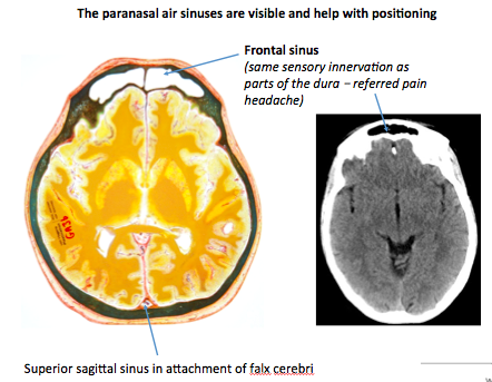
What sinus is shown?
what is its sensory innervation and how is this clinically relevant?
How does this help us to know level of positioning?
What sinus is shown posteriorly?
Frontal sinus
Sensory Innervation of the frontal sinus is the same as parts of the dura. Dura is innervated by different divisions of trigeminal (v1/v2/v3), frontal sinus is innervated by V1, therefore inflammation of the meninges can refer to the frontal sinus causing headache.
Aids positioning of the scan as frontal sinuses are within the frontal bone superior to the eyebrow.
Superior sagittal sinus is shown posteriorly, in attachment with the falx cerebri.
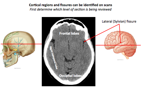
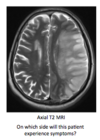
What lobes are shown?
What level is this scan at?
What fissure can be seen?
1) Frontal lobe
2) R temporal lobe
3) Occipital lobe
Fissure –> Sylvian or lateral fissure
Level of the scan must be at the level of the eyebrow region as the two frontal sinuses are shown.
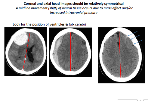
What should coronal and axial head images normally be?
Is this is not identified what pathology may be indicated?
What anatomical features can be used to indicate pathology in a head scan?
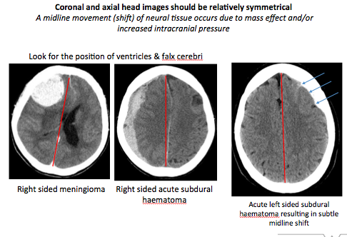
- Coronal and axial head scans should normally be symmetrical
- Midline shift indicates there is a space occupying lesions, or increased intracranial pressure
- Anatomical features that can be used are –> falx cerebri, position of the ventricles
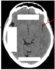
What pathologies are shown in the following images?
- Right sided meningioma with large left midline shift
- Right sided subdural haematoma with slight left midline shift
- Left sided extradural haematoma with slight right midline shift
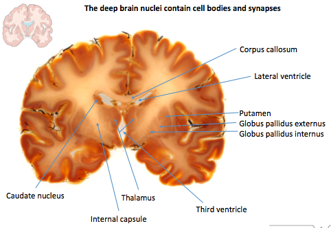
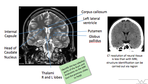
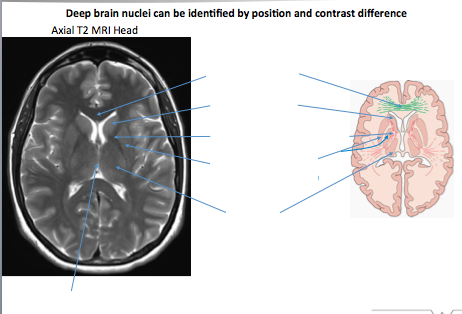
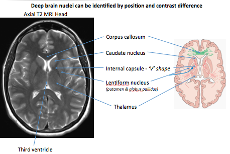
Label image shown
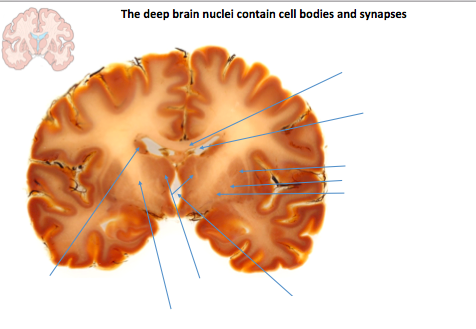
Top to bottom R to L:
- Corpus callosum –> connecting R and L hemispheres
- L lateral ventricle
- Putamen
- Globus pallidus externus and internus
- 3rd ventricle
- R and L thalamic lobes
- Internal capsule
- Head of the caudate nucleus
Label the image shown
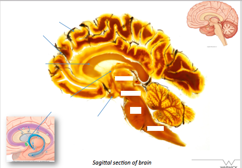
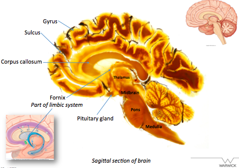
What is the thalamus composed of?
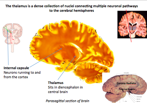
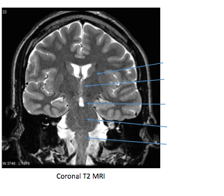
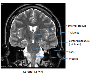
Label the image shown
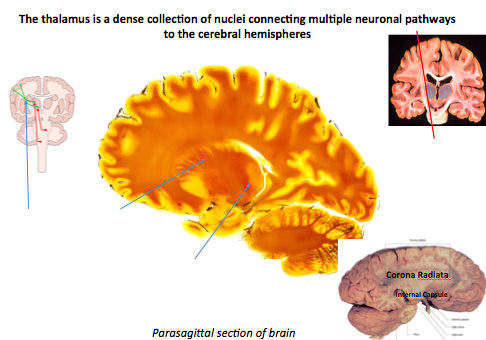
- The thalamus is a dense collection of nuclei that connects multiple neuronal pathways to the cerebral hemispheres
- The first arrow is to the fibres of the internal capsule running between the cerebral cortex and other brain regions/ spinal cord.
- Second arrow to the thalamus which is part of the diencephalon.
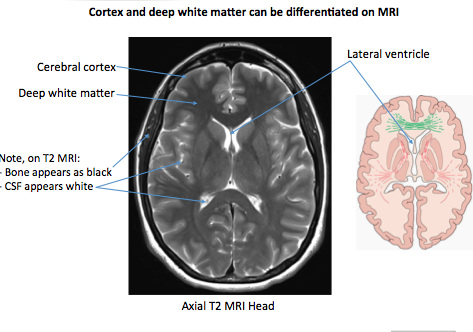
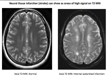
Label the image
T2 weighted MRI:
What is the appearance of bone?
What is the appearance of CSF?
On a T2 weighted MRI bone will appear black and CSF will appear white.
Note also that cerebral grey matter can be distinguished from white matter tracts on a T2 weighted MRI.
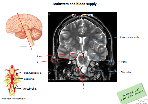
Label image
What are the arrows pointing to on the image?
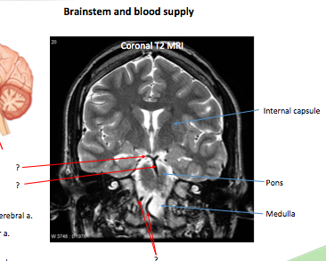
- Arrows pointing the blood vessels supplying the brain
- Bottom two arrows pointing to the vertebral arteries
- Then basilar
- Then posterior cerebral arteries
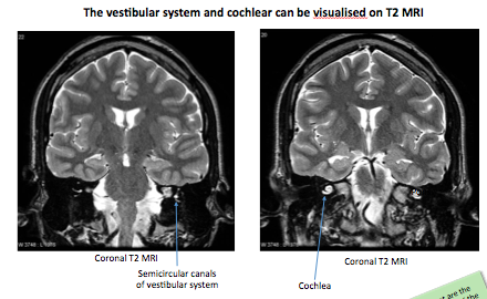
What two important structures are shown in the image?
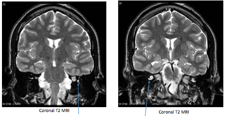
- Left hand image pointing to the vestibular system, the semicircular canals –> balance
- Right hand image is pointing to the cochlea –> contains inner hair cells that allow transduction of sound via auditory nerve to auditory cortex.
Label the image
How does neural tissue appear on a T2 MRI after a stroke?
What is a watershed infarct?
- Damaged tissue from a stroke will show with a higher intensity signal (brighter).
- Watershed infarct also known as a border zone infarct and occurs at the border of vascular territories, where tissue is farthest from arterial supply and therefore most vulnerable to reduction in perfusion.


-
Block 2 Week 2 Thorax/ Tracheobronchial tree/ lungs39
-
Block 2 Week 2 Thorax Tracheobronchial tree/ lungs part 251
-
Larynx34
-
Heart and mediastinum69
-
Cardiac embryology33
-
Vertebral Column41
-
Introduction to CNS anatomy53
-
Block 3: Spinal cord and ascending tracts45
-
Block 3: spinal cord and descending tracts / reflexes33
-
Block 3: Basal ganglia19
-
Cranial nerves48
-
Brainstem46
-
Cerebral cortex and limbic system49
-
Intro to neuroimaging35
-
Eyes- orbit, movement and reflexes33
-
Gluteal region, hip and thigh46
-
Intro to musculoskeletal27
-
Proximal neurovasculature, Knee and Leg39
-
palpable masses32
-
distal neurovasculature, ankle and foot47
-
Pectoral Girdle/ Should/Arm/Elbow48
-
Injuries to bones and joints of lower limb38
-
Upper Limb Forearm and elbow39
-
Wrist and hand36
-
Abdominal viscera: Liver, Pancreas and Spleen36
-
Female Reproductive Anatomy64
