What determines the smallest object we can see?
- size at which objects become visible depends on resolution of observers eye
*res = smallest distance between 2 objects that allows us to see them as seperate objects
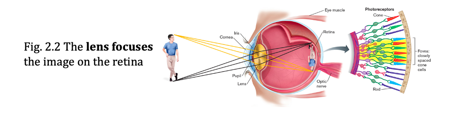
- resolution determined by the distance between 2 foveal pixels (group of cones with neurons in the portion of the retins where photoreceptors are packed)
- we can detect a group of smaller items when in a group but cannot resolve them as individuals
describe magnification
- need to magnify to resolve (inc the objects apparent size/dimension
- detection = ability to determine the presence of an object
*eukaryotes range from 10-100 um, prok range from 0.4-10 um
examples of size based contradictions of microbes
- some can be seen with the naked eye: Thiomargarita namibiensis
- some function as cell communities ex: biofilms (a whole community of bacteria either 1 species or several, we need to study as community)
- some microbial bacterial communities should be studies as whole entities: the human gut/microbiome
- some viruses are as large as bacteria: mimivirus, mamvirus
-
what are the different instruments to look at microbes
- light microscopy: resolves images according to absorption of light
- Electron microscopy: uses means of electrons to resolve smaller details (smaller then the wavelength of visible light, can resolve viruses)
*electron beams have smaller wavelength so can resolve smaller things
- atomic force microscopy: uses intermolecular force to map 3D topography of the cell
- X-ray crystalography: detects the interference patters of X-rays entering the crystal lattice of a molecule
what is the range of visible light? what is the conditions required for electromagnetic radiation to exist?
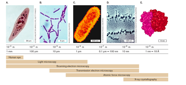
- visible light (400-700nm) is part of electromagnetic radiation (electrical and magnetic waves perpendiular to each other
- for electromag radiation to resolve an object the following must exist:
- wavelength of radiation much be equal or smaller than the size of the object (allows you to see that object as a seperate entity)
- there needs to be contrast between the object and its medium/background
- a detector with sufficient resolution for the given wavelength
*otpimal for 0.4-0.7 micro meters
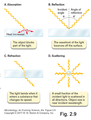
what are the 4 fates when light interacts with an object
- Absorption: photons energy is aquired by the absorbing object
*use for birght feild microscopy (object appears dark), in dark feild the object is very bright
- Reflection: the wavefront bounces off the surface of an object
- Bending of light as it enters a substance that slows it speed
- Scattering: the wavefront interacts with an object smaller than the wavelength of light
what are the 4 types of light microscopy
4 types:
Bright field microscopy
Dark field microscopy
Phase-contrast microscopy
Fluorescence microscopy
describe bright feild microscopy
- most common
*subcellular structures too small to resolve by light microscopy
- object appears as a dark silhouette
- resolution limit = 0.4-0.7 but magnification 1000x (greatest magnification that can imporve our preception of detail)
- greater magnification inc the image size but not the resolutions (empty magnification)
- can use oil emerson lens/microscopy
what is oil emersion lens microscopy
- use in birghtfeild
- put a drop of oil (similar refractive index to glass lens) between the lens and the object minimizes loss of refracted light at the widest angles and sharpens image (see Fig 2.14)
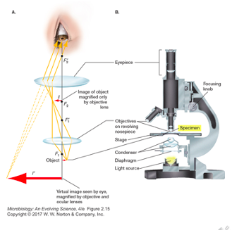
what is a compound microscope
- workhorse light microscope in gneral microbio labs
- has a system of multiple lenses designed to focus, correct &/or compensate for abberration
- ocular lens (10x magnification), objective elns (10x-400x mag) *needs to be parfocal)
- total magnification = magnification of the ocular multiplied by that of the objective
what is a simple way to observe microbes
- place them in a drop of water on a slide with a coverslip
- called wet mount prep
Advatages: observation of cells in natural state
Disadvantages: little contrast between cell and background (want to stain cells), sample may dry out qucik (use mounting reagents)
*we are using chemicals which are quite harsh (alc and acids), this kills the cell and we
*detection and resolution under a microscope are enhanced by staining, staining does NOT improve resolution
what is the difference between simple stain and differential stain
Simple stain
- adds dark colour specifically to cells, but not to external medium or surrounding tissue
- methylene blue is the most commonly used stain
Differential stain
- stains one kind of cell but not another
- most famous differential stain is gram stain
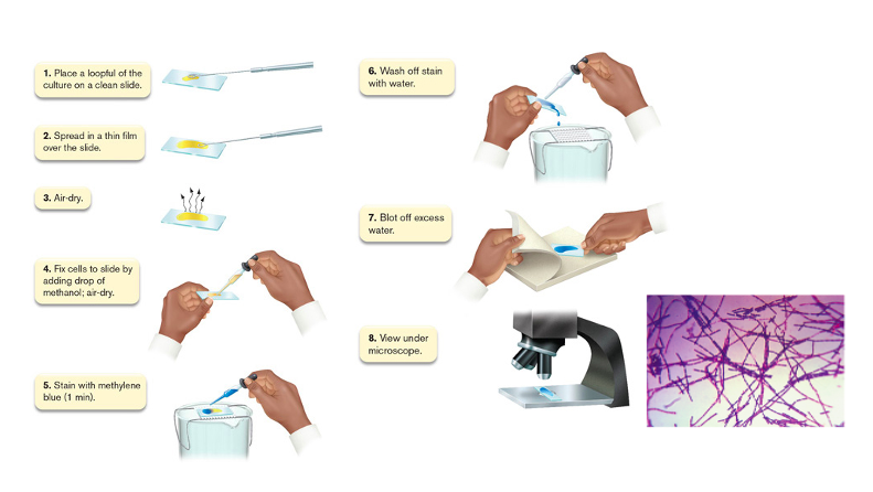
Describe the staining procedure for Methylene Blue
- methylene blue is a general stain for cells, just adds colour
1. put sample on slide with water so you can spread the sample
2. allow to air dry which may shrink the cells
3. add methanol to fix cells to the slide, allow to air dry
4. stain with methylene blue (1 min)
5. wash off stain with water
6, blot off excess water
- view under microscope
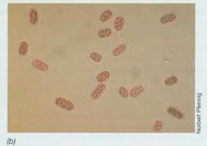
what is a differential stain, give an example and how it works
- stains one kind of cell but not another
- Gram stain differentiates between 2 types of bacteria
- Gram positive: retains the crystal violet stain because of thicker cell wall (cells appear purple)
- Gram negative: do not retain stain, cells appear pinkish/reddish
explain the Gram stain procedure
- Add methanol to fix cells to surface
- Add crystal violet stain
* stains gram pos cells reversibly - Add iodine which binds stain to Gram-pos cells
* only gram pos cells have iodine complex with crystal violet to retain the stain - Wash with ethanol
* stain is removed from gram neg but remains in gram pos - Add safranin counterstain
* counter stains gram neg pink, gram pos stay purple

explain the staining mechanism
- gram positive has multiple peptidoglycan layer
- gram neg have a thin peptidoclygan layer sandwhiched between two membranes
- when crystal violet it added it penetrated into the cell wall/ peptidoglycan (dye penetrates through all layes in gram pos)
- wash to remove excess
- by adding iodine, it complexes with the crystal violet and gets traped in peptidoglycan layers
- add the counter stain to stain cells that did not retain a lot of crystal violet

what is a spore stain?
- another differential stain
- uses malachite green, detects endospores of Bacillus and Clostridium bacteria
- retained by spores bc very different cell walls
cells highlighted as unstained halos
what is capsule staining
- also called negative staining
- its a differential stain
- does not stain the capsule, the halo you see is the spore around the bacteria cell
- stain interacts with background and cells highlighted as unstained halos
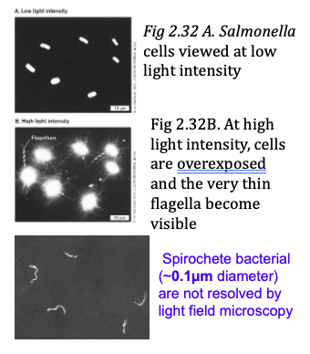
what is dark field microscopy?
- dark feild optics enables microbes to be visualized as halos of bright light against darkness
*visualize live samples bc no fixation or staining required
mechansim: object scatters light and is collected by objective lens, light that just passes through slide shines outside the lens so backgorund is dark
*important for cells that are very thin
*see outline of cels but not detail
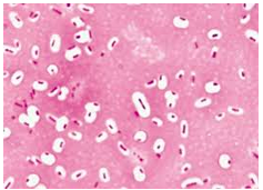
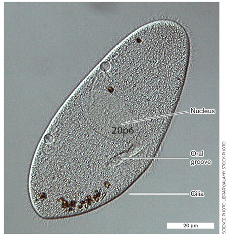
What is phase-constrast Microscopy?
- PCM
- expolits differences in refractive index between the cytoplasm and the surrounding medium or between diff organelles
- reveals differences in refractive index as patters of light and dark
*can be used to view live unfixed organelles, useful for eukaryotes cells protozoa and ameba
* in picture the differences in refractive index reveals the nucleus, oral groove and cilia
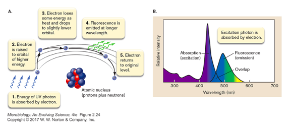
what is fluorescence microscopy
- powerful tool for detective ‘parts’ of cells
- specimen absorbs light as a defined wavelength and emits light of a longer wavelength plus some heat (fluorescence)
- scope fitted with light detectors, use different probes to detect components within cell after they have been labelled with a prob
- can visualize live (or fixed) cells in three dimensions
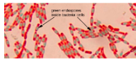
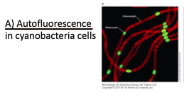
What is Autofluorescence?
some cell components naturally fluoresce under specific light wavelengths (no stain required)
* will fluoresce when light of a certain wavelength is shined
- cells with chlorophyl will autofluorece
ex: cyano bacteria
* in image the green is heterocyst which has been stained with a specific probe, the cells containing chlorophyl are autoflurorescing as red
what are fluorophores?
- tags that are attached to a probe that interact with specific components of a cell
*fluorescent chemical compound with specificity for cellular target
- this specificity is determined by: chemical affinity, labelled antibody, DNA hybridization, Gene fusion reporter tags (when rpotein is expressed you see where is it located in cell)
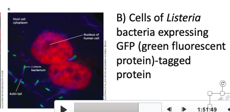
what is GFP
•Jelly fish Green Fluorescent protein (GFP)
- the DNA sqeuences coding for GFP can be spliced to that of a target protein gene sequence (to create a recombinant gene)
- this is then expressed into a protein (which is fluorescent bc of the GFP-tag)
* tag a specific component and you see that, you can also tag multiple components


-
History of Microbiology30
-
Microscopy31
-
Bacterial CM transport29
-
Cell envelopes35
-
Virus37
-
Microbial growth30
-
Biofilms18
-
Environmental influences on microbial growth25
-
Microbial diversity16
-
Bacterial diversity27
-
Archaeal Diversity24
-
Microbial Diversity: Eukarya33
-
Microbial Ecology and Microbial Association46
-
Introduction to the innate immune system35
-
Cellular barriers to the innate immune system33
-
Adaptive Immune system27
-
Adaptive Immune system Pt 2: Oct 3021
-
Guest lecture19
-
Microbes in health and disease31
-
Microbes and health: Nov 625
-
Defining the interplay of Klebsiella pneumoniae and host during infection by quantitative proteomics24
-
Viral Pathogens26
-
HIV: Viral pathogen24
-
Guest lecture Nov 1812
-
Physical, chemical and biological control of microbes38
-
Microbes in biocontrol19
-
microbes in food and beverage industry17
-
lab11
-
microbes in food and beverage industry pt 223
