What key areas should you review on CXR?
- Lung apicies
- Heart
- Diaphragm
- Bones & soft tissue
- Lines/wires
Name some main visible structures on a CXR
- Trachea
- Hila
- Lungs
- Diaphragm
- Heart
- Aortic knuckle
- Ribs
- Scapulae
- Breasts
- Bowel gas
Name some importnant obscured structures on CXR
- Sternum
- Oesophagus
- Spine
- Pleura
- Fissures
- Aorta
What shape should the aortopulmonary window be?
What should you consider if it is not that shape?
Normally: concave
If straightened or convex consdier Mediastinal lymphadenopathy
Name some structures in the AP window
- Left phrenic nerve
- Left recurrent laryngeal nerve
- Left vagus nerve
- Left bronchial arteries
- Ligamentum arteriosum
- Fat
- Lymph nodes
Consolidaiton VS Collapse
Consolidation: Alveolar airspaces filled with fluid/ tissue/other matrial
Lobar collapse: Signs of volume loss & absences of air bronchograms
You may see the fissues and hilar having moved up or down
Which lung hilum is normally higher
left lung hilum
What is (loss of) silhouette sign indicate? (very broad)
Pathology

What imaging features should you look for on CONSOLIDATION?
A2BC3
- Acinar rosettes- fluffy appearance of parenchyma distal to terminal bronciole
- Air bronchograms (tubular outline of airways made visible due to alveolar filling of fluid/ inflammatory exudates)
- Bat wing distibution
- Confluent ill-defined appearance
- Consolidation
- Change occurs rapidly
PA CXR
What is this showing? Collapse or consolidation?

Consolidation. Hilar are equal levels. Ill defined appearance
What is this showing?
AP errect

LUL collapse- collapses anteirorly becoming a thin sheet of tissue –> “Veil sign”
Loss of silhouette sign of aortic knuckle
General opacity on the left
What is this AP CXR showing?

Left Lower Lobe collapse –> Sail sign
Loss of silouette sign of left demidiaphragm and descending aorta
Inferior displacement of left hilum
Inferior displacement of oblique fissue
What is the PA CXR showing?

Right middle lobe collapse. Child has inhaled foreign body
Loss of horizontal fissure
Loss of silhouette sign of right hear boarder
What is this PA CXR showing?
What should you be suspicious of?

Right upper lobe collapse –> Golden S sign
Consider: carcionma. The mass can block the bronchus to right upper lobe causing it to collapse and the lung rotates backwards

What should you never XRAY if you have suspicions of?
What is the treatment for this?
Tension pneumothorax
Tx: 14G cannular –> 2nd ICS MCL
For an ETT placement what are the rules of placement
- Assess mandible position: flexed, extended or neutraul (over C5/6)
- Desired position when neck is neutral: 5+/-2cm above carina
Flexed neck- 3cm (+/- 2 cm)
Extended neck- 7cm (+/- 2cm)
If incorrectly places can cause right lung hyperinflation and left lung collapse
What are the rules of NG tube placement?
- Descends thorax in midline
- Bisects carina
- Crosses diaphragm in midline
- Tip sits below diaphragm

What is this HRCT showing?
What is it indicative of?
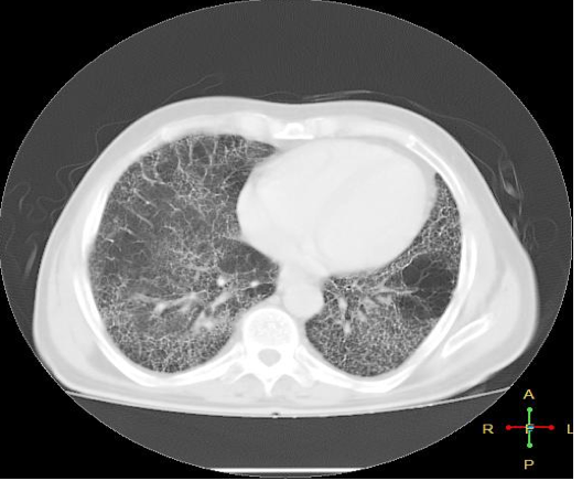
HONEYCOMBING indicative of pulmonary fibrosis
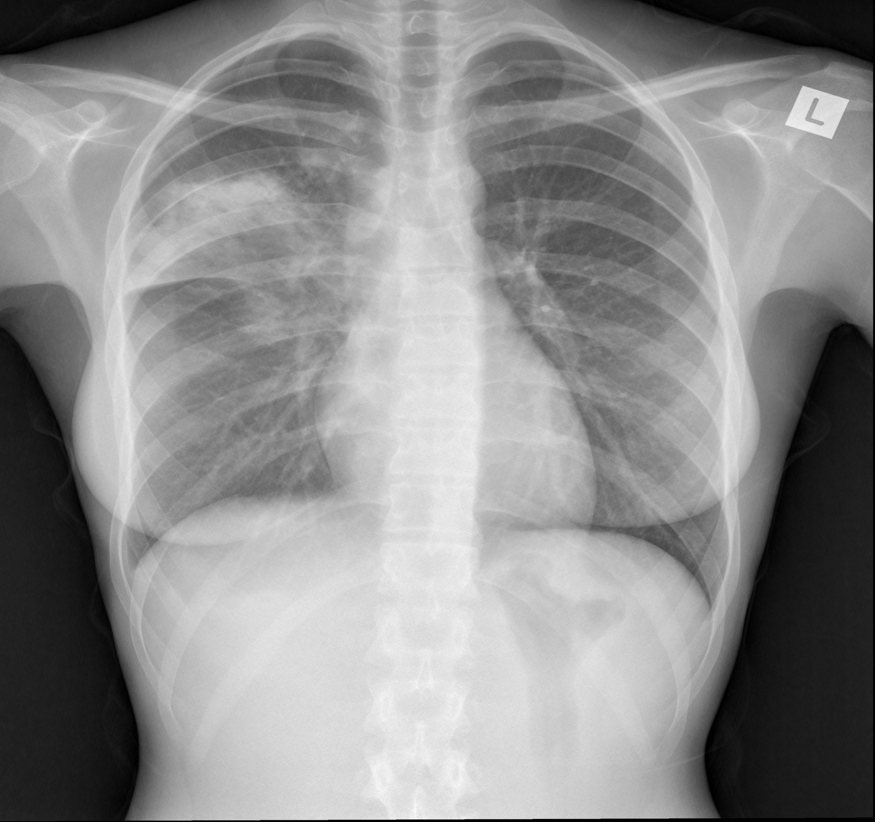
What is the CXR showing?

Reticular shadowing- looks course, nodular type pattern unlike consolidation/ pulmonary oedema
“Reticular nodular” is typical of a fibrotic lung condition
Then do high resolution CT scan
What is this CTPA showing?

Pulmonary embolism. Areas of radiolucency are the PE
What blood test should you never do in pregnant women who you suspect have a pulmonary embolism
D-Dimer. Already in a hyper-coaguable state
What is this CXR showing?

Radio opacity and miniscus present thus PLEURAL EFFUSION
-
Psychology55
-
Anatomy295
-
Week 1- AMC lecture33
-
Histology17
-
Week 2-Neck anatomy 157
-
Week 2-Neck anatomy 250
-
Week 2-Psychiatric history33
-
Week 2-Mental state examination24
-
Week 2- Definitions MH31
-
Week 2- Advance Decisions to Refuse Treatment and Advance Care Planning19
-
Week 2- Cardiac & Vascular imaging20
-
Week 3- Venous and arterial access47
-
Week 3-Lumbar puncture, epidural, spinal and caudal47
-
Week 3- Complexity in Human Behaviour13
-
Week 3-UTI18
-
Week 4- Thorax imaging22
-
Week 4- Asylum seekers15
-
Week 4- Inequities in health care16
-
Week 4- Homelessness & Health16
-
Week 7-DM25
-
Week 7- Concordance34
-
Week 7- Children & refusal of consent21
-
Week 8- children & young people with life limiting illness12
-
Week 8- Medical Imaging Abdomen23
-
Week 8-paediatric inherited conditions29
-
Week 8- Epigenetics and Cancer19
-
Week 9-Neoplasia 136
-
Week 9-molecular cell biology of cancer23
-
Week 10- Career psychology notes2
