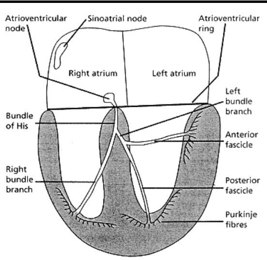
how does the conduction system of the heart work
- generates its own electrical activity which is conducted via a specialized set of cells from the atria to the ventricles
- eletrical activity of this conduction system is passed to the muscle cells (cardiomyocytes) which then contract via the process of excitation-contraction coupling
1. SA node generates spontaneously depolarizing cells (pacemaker cells)
2. when they fire the conduction passes from the right atrium to the left atrium and the AV node
3. the AV node has pacemaker cells but they are slower –> down to the Bundle of His
4. conduction reaches the purkinje fibres
5. passes to outer wall of the heart (epicardium)
6. occurs every half beat
what is the dipole concept
- an electrical dipole is a negative and positive charge separated by a distance
- dipole creates a voltage
- voltage can be measured using a voltmeter
- voltmeter has two electrodes one denoted as (+) and the other (-)
- if positive end of the dipole is nearer the (+) electrode then an upward deflection in the voltage trace occurs (vice versa)
what is the resting membrane potential of a cardiomyocte
-80mV
inside is negative and outside is positive
what occurs during depolarization in a cardiomyocyte
positive ions enter the cardiomyocte –> inside becomes positive and the outside becomes negative
what occurs during repolarization
interior of cardiomyocyte becomes negative again and outside becomes positive
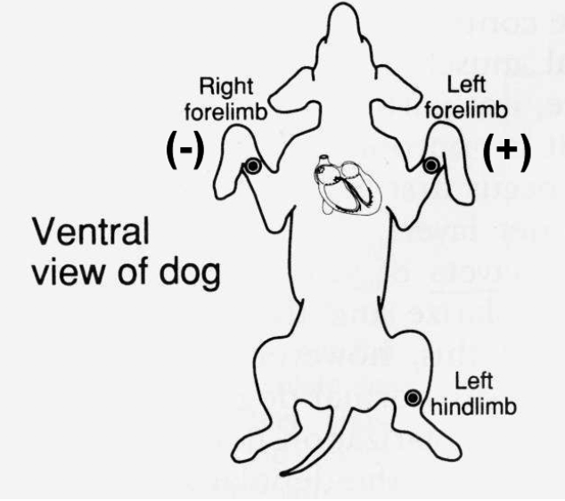
explain the dipole concept in the whole heart (5)
- volmeter is the ECG machine and the electrodes are placed on the body
- ECG machine measures the changes in voltage created by depolarization and repolarization of the different regions of the heart during the cardiac cycle
- one electrode is placed on the left forelimb and one on right
- ECG is record of voltage in the left forelimb with respect to the right therefore the left forelimb is denoted as the (+) electrode
- if a relatively more positive region of the heart is closer to the left electrode an upward deflection of the ECG trace will result (vice versa)
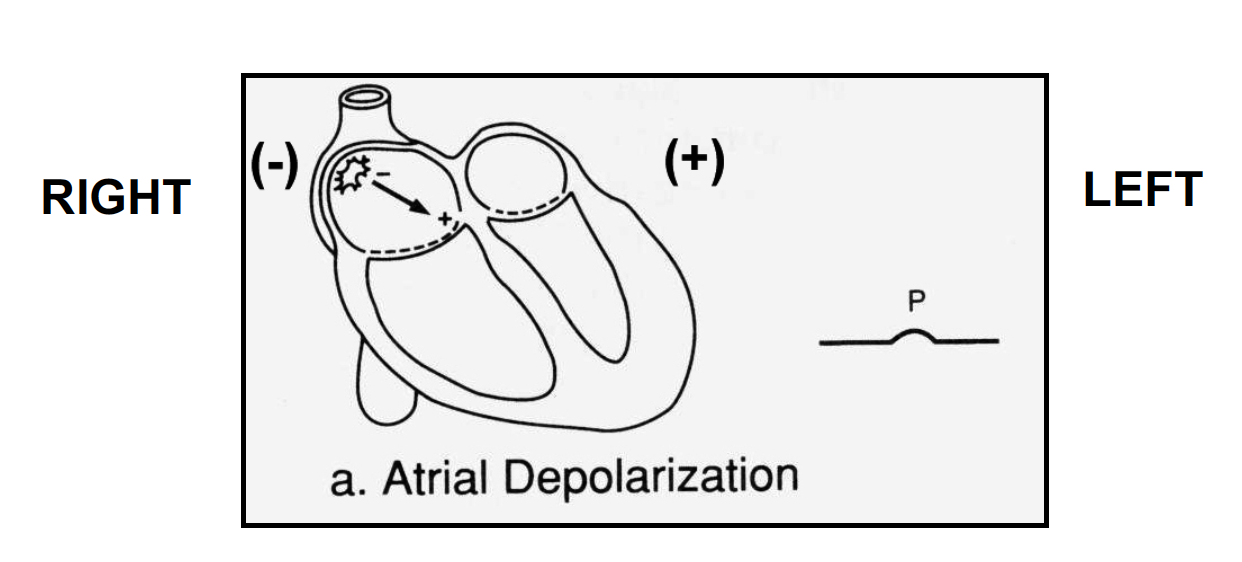
explain what occurs in atrial depolarization and what will appear on the ECG trace (6)
- the SA node cells in the RA spontaneously depolarize and result in depolarization of the adjacent RA cells
- the wave of depolarization moves towards the left as demonstrated by the arrow
- the cells in the LA are at rest hence a dipole is created
- positive end of the dipole is closer to the (+) electrode hence an upward deflection occurs in ECG trace
- when all the RA and LA is depolarized the trace returns to base-line
- next delay is the wave of depolarization passing through the AV node to ventricles
explain what occurs during early ventricular depolarization
- wave of depolarization passes down interventricular septum
- wave of depolarization spreads from left to right creating a dipole
- the negative end of the dipole is closer to the (+) electrode hence a downward deflection in the ECG trace occurs
*left hand side of septum depolarizes first so dipole –> negative end closer to the voltmeter –> trace goes down whole septum depolarizes and trace goes to 0 –> early ventricular depolarization (Q wave)
what occurs during ventricular depolarization (4)
- endocardium depolarizes before the epicardium
- a dipole is created which is very large since the number of cardiomyocytes within the left ventricle are numerous
- the positive end of the dipole is closer to the (+) electrode hence a upward deflection in the ECG trace occurs
- a dipole is directed towards the left since –> the ventricular apex is directed towards the left and the left ventric is much larger than the right and therefore dominates electrically
**
once its gone thru septum –> goes down bundle branches to the purkinje bundles –> the outside starts to depolarize –> dipole positive end is closer to the positive end –> trace end goes up –> very high because greater number of dipoles created compared to the atrium (R wave)
the left apex is closer the left side of the chest (apex beat)
this has direction and magnitude –> vector
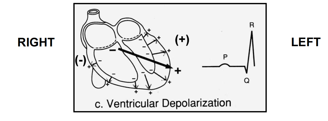
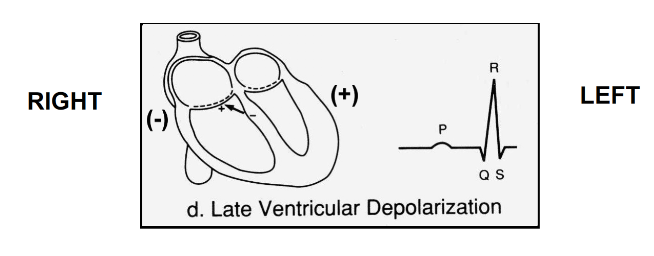
explain what occurs in late ventricular depolarization and what it appears like on the ECG
- wave of depolarization finishes spreading from the endocardium to the epicardium of both ventricles
- the ECG returns to the baseline point and sometimes goes negative
- the negative end of the dipole is closer to the (+) electrode hence a downward deflection in ECG trace
S wave: small negative deflection –> negative end is closer to the positive end
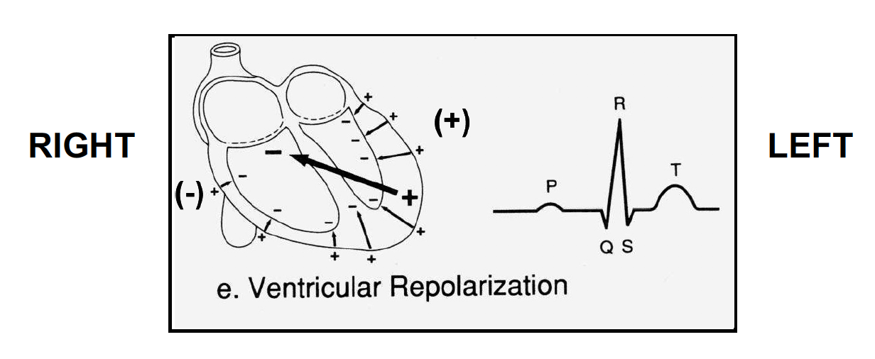
explain what occurs in ventricular repolarization and what it appears on the ECG
- the epicardium is the last to depolarize but the first to repolarize
- epicardial cells are now positive on the extracellular surface and create a dipole with endocardial cells which are still depolarized
- positive end of the dipole is closer to the (+) electrode hence a upward deflection in the ECG trace occurs
**the first cells to repolarize are on the outer surface of the myocardium –> cells go back to negative and the outside is positive –> closer to the positive electrode –> trace goes up even though its repolarization –> eventually whole thing repolarizes and the dipole is lost
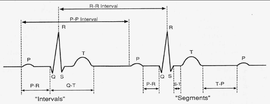
what is the P wave
atrial depolarization
what is the QRS complex
ventricular depolarization
what is the T wave
ventricular repolarization

what is the PR interval
time between atrial depolarization and ventricular depolarization

what is the QT interval
length of time that the ventricles remain depolarized

what is the QRS complex
time taken for ventricular depolarization to occur once the wave of depolarization has passed through the AVN from the atria

what is the PP interval
time between atrial depolarizations

what is the RR interval
time between ventricular depolarization
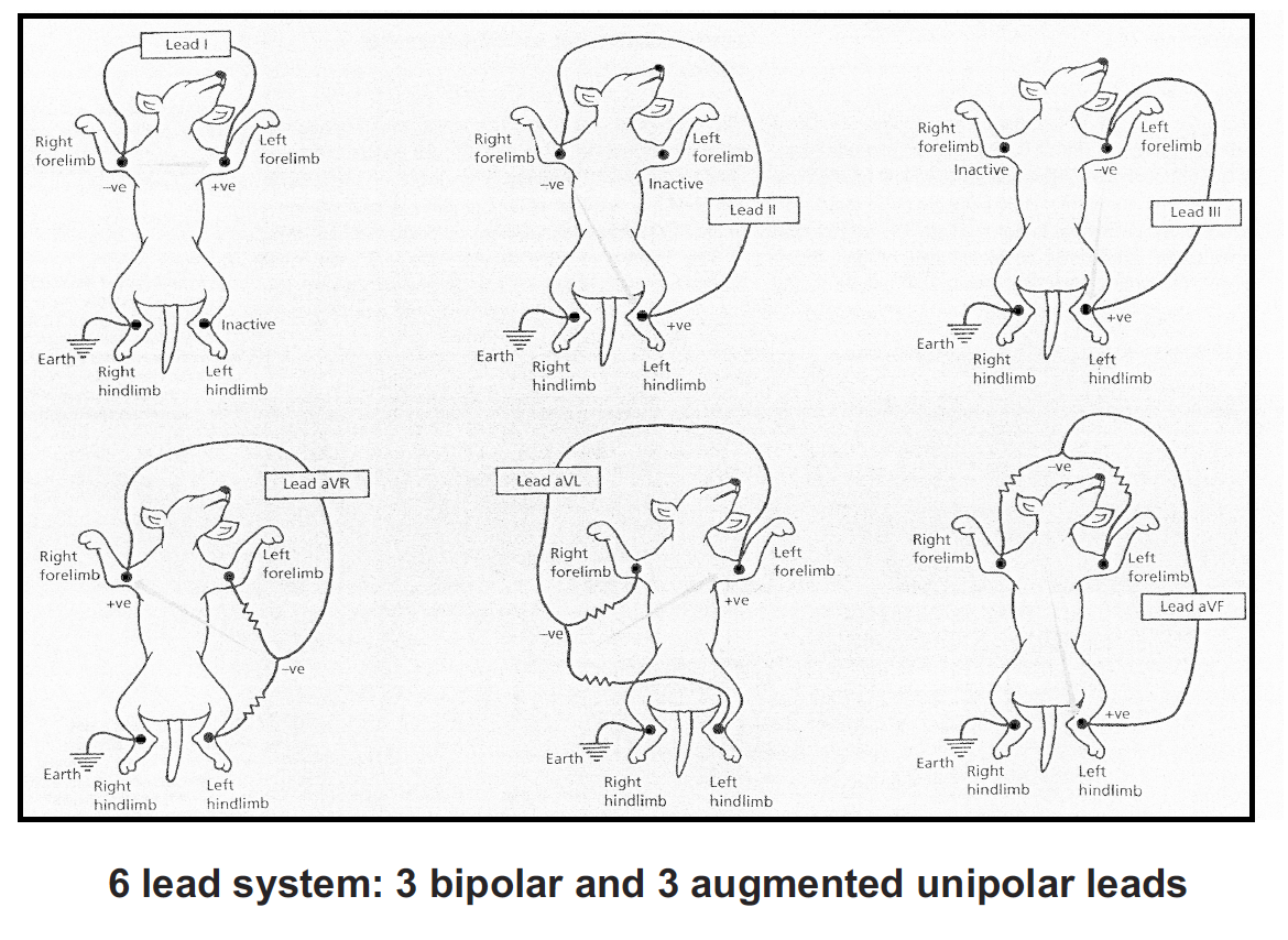
what are the 3 lead systems
lead I: right forelimb (-), left forelimb (+)
lead II: right forelimb (-), left hindlimb (+)
lead III: left forelimb (-), left hindlimb (+)
what are the clinical significances of ECGs
- heart rate (HR)
- normal sinus rhythm –> dysrhythmia (arrhythmia) means an abnormal rhythm
- chamber enlargement
- myocardial ischemia
- electrolyte imbalance
how is the heart rate calculated using ECGs
RR interval –> becomes shorter when heart rate increases (tachycardia)
how can arrhythmias be detected using ECGs
normal heartbeat initiates in the SAN since the cells in that region normally have the fastest intrinsic pacemaker activity
a dysrhythmia means abnormal rhythm
how can ECG be used to determine chamber enlargement
using the lead system a 3D appreciation of the eletrical activity of the heart is acquired
can be used to determine chamber enlargement (not horse, but dog)
**
vector quantity –> the R wave is very tall because it has more muscle compared to atrium
if the atria are enlarged there may be a change in the P wave
can tell this in the dog but not horse


-
Anatomy of Bloodflow Through Heart 136
-
Anatomy of Bloodflow Through Fetal Heart33
-
Normal Cardiac Electrical Function29
-
Normal Cardiac Mechanical Function11
-
Factors Affecting Cardiac Output38
-
Histology of Blood Vessels: Structure & Function47
-
Infectious Aetiologies of Heart (lungworms & heartworms)30
-
Diagnostic Imaging of Heart57
-
Branching of Aorta 153
-
Branching of Abdominal Aorta & Origins of Vena Cava & Azygous Veins22
-
Cardiovascular Pathology 159
-
Control of Blood Vessel Function33
-
Equine Vet & CVS33
-
Infectious Aetiologies of Heart (Bacterial + Viral)33
-
Cardiovascular Pathology 268
-
Homeostasis49
-
Cardiovascular Related Physiological Adaptation to Disease37
-
Cardiovascular Pathology 357
-
Pathology of Vessels56
-
Pharmacological Options in Heart Failure86
-
Antiarrhythmic Drugs49
-
Heart Dissection64
-
Cardio & Resp Histology59
-
Ultrasound of Heart SDL48
-
Thorax Practical81
-
Radiography of Heart SDL23
-
ECG Practical4
