Physiology1 Flashcards

1
Q

Action potential graphs comparison between atrium, ventricle and SA node.

A

Atrium reaches peak quickly and drops with no plateau but a slow drop, ventricle peaks very shortly after followed by a plateau, SA node peaks first with an equal rise and drop.

2
Q

SA node action potential

A

The nodal cell action potential is easy to recognize since there is no plateau phase, the depolarization phase is slower than in the atrial or ventricular cell and the resting membrane potential is achieved briefly as the cell continues to depolarize automatically until the next action potential is triggered.

3
Q

Divisions of the action potential in the ventricular cell

A

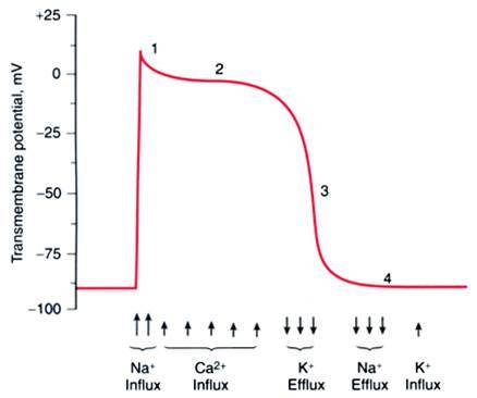
Phase 0 is the rapid depolarization, phase 1 is a transient repolarization, phase 2 is the plateau phase where the contraction happens, phase 3 is the repolarization phase and phase 4 is the resting potential phase.

4
Q

Membrane potential regarding ion influx in phases of action potential in a fast fiber

A
5
Q

Graph of the channels in slow fiber action potential

A
6
Q

Phase 0 of the fast fiber action potential

A
  1. at -90mv, gates closed but small chemical gradient and large electrical gradient, 2. at -65mv, gate is open and Na+ runs through following electrical and chemical gradient. 3. At 0mv, no electrical gradient but Na follows chemical gradient, 4. at +20mv it continues to follow the chemical gradient but is opposed by the electrical gradient, 5. at +30mv the channel is closed and Na+ can’t enter because the electrical gradient overpowers the chemical gradient.
7
Q

What are the two sodium channel gates?

A

m (activation gates) and h (inactivation gates).

8
Q

Where does transient outward current occur?

A

Phase 1

9
Q

Where does the Delayed rectifier current occur?

A

Phase 1 and 3

10
Q

Where does the inwardly rectified current (Ikl) occur?

A

Phase 1 and 4, during repolarization from -20 to -70mv

11
Q

What channels control the small repolarization in phase one

A

Ito channels

12
Q

What potassium channels/currents are active in phase 2?

A

All (Ito, Ik, Ikl) but overpowered by Ca2+ influx and potential stays the same.

13
Q

What channels are active in phase 3?

A

Most effect of Ik. All 3 (Ito, Ik, Ikl) to counteract Ca2+ influx and repolarize cell.

14
Q

When are L Ca2+ channels open?

A

+10mv, during phase 0 of the fast fiber action potential, open rapidly but deactivate slowly

15
Q

When are T Ca2+ channels open

A

-20mv, during phase 0 of the fast fiber action potential, before the L type

16
Q

Which are the are the most abundant of the Ca++ channels?

A

L type

17
Q

The — of Ca2+ balances out the — of K+ to maintain the membrane potential unchanged and cause the plateau phase of the fast fiber action potential

A

influx, efflux

18
Q

What ends the plateau phase and begins repolarization?

A

The Ca++ channels close and K+ efflux predominates

19
Q

What is the effect of calcium blockers (Diltiazem)?

A

L type

20
Q

What is the effect of Diltiazem (calcium blocker)

A

Decreases plateau phase, lets K+ currents have an upper hand so that repolarization in phase 3 begins much earlier. Starts to make ventricle AP look like atrium.

21
Q

Torsades de Pointes Phenomenon/Early After Depolarizations

A

Lack of initiation of phase 3 due to failure of calcium ions responsible for depolarization. Na and Ca channels are not slowed and depolarization is repeated without allowing K to flow out and repolarize. Need to shock.

22
Q

What causes Early After Depolarization

A

Calcium continuously entering.

23
Q

What are fast Na+ channels?

A

Phase 0 depolarization of non-pacemaker cardiac action potentials (Atria, ventricles, Purkinje)

24
Q

What are slow Na+ channels?

A

“Funny” pacemaker current (If) in cardiac nodal tissue (SA, AV)

25
Q

Action potential graph of a slow response fiber

A
26
Q

What are the main characteristics of the slow fiber action potential?

A

A slower phase 0 in which Ca++ becomes an important component of the depolarizing current, no phase 1 and very little or absent phase 2.

27
Q

Relative Refractory Period (RRP)

A

This means that the fiber will not respond with a full amplitude action potential until it is well into phase 4.This makes the fiber less responsive for sometime even when it is in phase 4 and creates a period of extended refractoriness.

28
Q

Gap junctions role in action potentials

A

These junctions create an electrical coupling among all the fibers and greatly promote conduction. Remember that in the cell local current refers to the movement of ions. Thus between two fibers you can see a current of positive ions moving across the gap junctions from fiber A to B intracellularly and depolarizing fiber B. Extracellularly, they move from B to A. The original depolarization in A was caused by an action potential that originated in this fiber or an adjacent one.

29
Q

What is the effect of high EC K+ on AP of fast fibers?

A

Decreased slope and amplitude. Eventually looks like a slow fiber. Eventually flatlines. Create a block and generate an aberrant cardiac rhythm by interfering with normal pacemaker.

30
Q

How is coronary disease related to high EC K+?

A

Causes low blood flow around the heart and diminishes activity of Na+/K+ ATPase in the heart cells. K+ accumulates extracellularly and causes decreased AP.

31
Q

What is the mechanism in which high K+ affect the voltage gates and the AP?

A

Lowers membrane potential, closes some h gates, inhibits Na+ entry, lowers AP amplitude.

32
Q

What are the ranges of K+ that the body can tolerate?

A

2.5-6.5 mEq

33
Q

Do fast fibers have a refractory period?

A

Yes, but all the fast Na+ channels are totally recovered at the end of phase 3. Complete response in phase 4.

34
Q

Post Repolarization Refractoriness

A

Period of prolonged refractoriness, conducts more slowly during this period and is more prone to conduction blocks.

35
Q

Why is a stimulation in early RRP of a fast fiber similar to a slow fiber AP?

A

Because there is high K+ extracellularly and this blocks the h gates from allowing Na+ entry.

36
Q

How long is the delay from the onset of phase 4 to the time in which a slow fiber will respond again with a complete AP?

A

200ms

37
Q

What happens if the SA node is damaged by an infarct in that area?

A

The Atrioventricular Node (AV) takes over until eventually the atrial cells develop another pacemaker. In addition there is a safeguard system formed by the idioventricular pacemakers (Ips) that are capable of firing at a reduced rate of 30 to 40 beats per minute.

38
Q

What are idioventricular pacemakers (Ips)?

A

Safeguard system. Ventricular conduction fibers that take the role of pacemakers. This usually happens when the AV node cannot conduct the impulse coming from the SA node to the ventricles.

39
Q

What is the pacemaker potential (PaP) period?

A

A period in which the cell depolarizes automatically. During this time there is a slow depolarization that gradually brings the cell to a threshold potential beyond which an action potential occurs. In reality the pacemaker potential is happening during phase 4 of the action potential.

40
Q

How does the PaP generate?

A

The genesis of the pacemaker potential (PaP) invloves three ionic currents. During the early period of the PaP a small Na+ (If) current starts to depolarize the cell. This current is followed by a large inward Ca++(iCa) current that eventually brings the membrane potential to the threshold and is also the main current carrying the positive charge inside the cell during the depolarization phase.

41
Q

What is the role of the K+ current in the pacemaker AP?

A

Influx is lower during the pacemaker potential phase but efflux increases dramatically at the peak of the depolarization phase and is the main current involved in bringing the cell membrane potential back to the minimum repolarization potential.

42
Q

What is the effect of catecholamines or cholinergics on the pacemaker potential?

A

Catecholamines hasten the pacemaker potential by increasing the slope. Cholinergics decrease the slope, slowing the pacemaker potential.

43
Q

Which has more of a basal effect on heart rate, catecholamines or cholinergics?

A

Cholinergics. HR increases when the heart is removed from the body (denervated).

44
Q

How do catecholamines act on the pacemaker action potential?

A

Catecholamines like epinephrine and norepinephrine act by increasing the Ca+ and Na+ currents and decreasing the K+ current involved in the pacemaker action potential.

45
Q

What is the effect of cholinergic stimulation on the pacemaker potential?

A

Cholinergic stimulation via the release of acetylcholine brings about a depression of the if and iCa currents and increased K+ current. The increase in the K+ current is mediated by specific receptor operated K+ channels that open in response to acetylcholine and hyperpolarize the cell.

46
Q

What is the effect of L type channel antagonists like Nifedipine on the AP of pacemaker cells?

A

lowers the amplitude of the pacemaker cell AP and also lowers the slope of the pacemaker potential reducing the firing frequency.

47
Q

What is overdrive suppression?

A

Explains why the area of the heart that fires with the greatest frequency is the one that regulates the heart beat. A greater firing frequency depolarizes adjacent cells by allowing Na+ to enter more frequently. Eventually the Na+/K+ ATPase is activated by the increased level of cytoplasmic Na++ created by the repeated rapid firing.

48
Q

How is an ectopic focus induced by coronary artery disease?>

A

High intracellular sodium causes activation of the Na+/K+ pump. Since the Na+/K+ ATPase is electrogenic and extrudes more Na+ in exchange for K+, this activation induces cell membrane hyperpolarization and increases the time required for depolarization thus suppressing automaticity in adjacent cells. It can fire at a frequency which is much higher than that of the SA node. If the ectopic focus suddenly stops firing, the SA node may be left momentarily suppressed causing the heart to stop and inducing syncope.

49
Q

What is Bachmann’s Bundle?

A

Fibers that conduct from the right to the left atrium. Posterior internodal pathways conduct from the SA to the AV node

50
Q

Nodal Delay

A

N region includes round cells that are slow conductors. Not only involves the slow conductors of the N region in the AV node but also the NA region where although conduction is rather fast the stimulus must transverse a longer path. As the stimulus approaches the N region it is slowed by the appearance of more round cells until the stimulus finds itself in the N region. NH region picks up again to the bundles. It can be extended by conditions that increase the conduction time through the AV node. Viewed as increased P-R interval in EKG.

51
Q

Mechanism of blockage of a bundle branch leading to a Paroxysmal Supraventricular Tachycardia: Bidirectinoal block of fast fiber

A

The AP will propagate through the fiber until the branch but if the fast fiber is blocked will only go through the slow fiber and this will compensate, albeit at a slower rate. No retrograde nor antegrade conduction through the fast fiber.

52
Q

Mechanism of blockage of a bundle branch leading to a Paroxysmal Supraventricular Tachycardia: unidirectinoal block of fast fiber causing Paroxysmal Supraventricular Tachycardia

A

Ischemia in fast fiber is depolarizable so AP traveling down can travel retrograde not antegrade through fast fiber, creating a counterclockwise reentry loop. This causes more depolarization in the slow fiber and in the whole ventricular region, creating an ectopic pacemaker.

53
Q

Pictures of bidirectional and unidirectional blocks of AP

A
54
Q
A
55
Q
A
56
Q

First Degree AV Block

A

Delayed conduction through AV node and EKG shows a P-R interval greater than .20 s. N cells show increased repolarization refractoriness which means that any stimulus falling in the PRR period of the cells will not be conducted. So, if the SA node fires at a very high frequency, conduction in the AV node is slowed. AV node act like a filter. Sinus rhythm and rate except elongated PR. Usually temporary problem (fever/infection).

57
Q

Second degree AV block

A

Frequency of atrial firing is so high that only half of the atrial stimuli make it through the AV node (1/2-4 atrial APs). Inverted T wave. No distance between P and T. Type I: PR interval starts normal and gets progressively longer, skips one and starts over. Type II: Extra P with missing QRS. Must be careful and possibly insert pacemaker, dangerous if reaches 3rd degree.

58
Q

Third Degree AV block

A

Sufficient vagal stimulation to completely block conduction through AV node. Ventricles are driven slowly by bundle of His or Purkinjes. P waves unrelated to QRS waves.

59
Q

Purkinje fiber size and structure

A

Specialized cardiac cells much larger than myocytes. Their diameter coupled to surface membrane specializations like lack of T tubules allows them to conduct very rapidly. Have a long Absolute Refractory Period (ARP), the slower the HR the longer the ARP. Stop-gap measure in case the AV node fires too quickly to keep cardiac rhythm in check.

60
Q

Purposes on an EKG

A

A recording of the electrical activity of the heart over time Gold standard for diagnosis of cardiac arrhythmias Helps detect electrolyte disturbances (hyper- & hypokalemia) Allows for detection of conduction abnormalities Screening tool for ischemic heart disease during stress tests Helpful with non-cardiac diseases (e.g. pulmonary embolism or hypothermia.

61
Q

Graph corresponding AKG to contraction and AP of myocytes in ventricles

A
62
Q

What is the size of one small block of ECG paper

A

1mm^2

63
Q

What is the time of one small block?

A

0.04s

64
Q

How much time is in one large block of ECG graph paper?

A

0.20s

65
Q

How many ECG blocks are there in a second?

A

5

66
Q

What is the voltage height of a small block?

A

0.1mV

67
Q

Normal conduction pathway

A

SA node -> atrial muscle -> AV node -> bundle of His -> Left and Right Bundle Branches -> Ventricular muscle

68
Q
A
69
Q

Intervals vs. Segments on ECG

A

From the end of a wave to the beginning of the next wave is a segment (end of P wave to beginning of QRS is the segment. ST segment is between the end of the S drop and the T rise), the isoelectic line portion. Intervals include a wave

70
Q

What is the cardiac cause of the P wave?

A

Atrial depolarization and contraction

71
Q

What is the cause of the QRS complex?

A

Ventricular depolarization

72
Q

What is the cause of the T wave?

A

Ventricular repolarization

73
Q

What does a prolonged PR interval indicate?

A

A 1st degree heart block

74
Q

What is an abnormal Q wave?

A

Greater than 1/3 R wave height, >0.04s. May represent MI.

75
Q

What is the normal duratino of a QRS complex?

A

0.08-0.12s

76
Q

Why would the P wave be absent?

A

No SA involvement.

77
Q

What is a nomal length of a P wave?

A
78
Q

What is the normal length of an ST segment

A

0.08-0.12s

79
Q

What is the interval from the beginning of the QRS to the apex of the T wave?

A

The absolute refractory period.

80
Q

QT interval

A

Measured from beginning of QRS to the end of the T wave. Contains ST segment and QRS. Normally about 0.4s but can get shorter with HR (exercise).

81
Q

What may increase the P wave?

A

Atrial enlargement (from mitral valve regurgitation)

82
Q

What is the normal duration of a PR interval?

A

0.12-.2s

83
Q

What will be the ECG effect during ischemic heart disease?

A

Depressed ST segment

84
Q

Sing bradycardia/tachycardia

A

Same sinus rhythm but rate <60 or >100.

85
Q

Flutter

A

Contraction rates 200-300/min. Can’t get through AV node-cut off. Sawtooth pattern with no isoelectric line. QRS gets through occasionally. Caused by reentry loops.

86
Q

Atrial Fibrillation

A

Contraction of myocardial cells uncoordinated and pumping ineffective. Life threatening. Electrical defibrillation resynchronizes heart by depolarizing all cells at once.

87
Q

Ventricular fibrillation

A

Coarse fib vs fine fib (looks like asystole).

88
Q

ar fibrillation and pulseless ventricular tachycardia.Defibrillation use corrects:

A

Arrhtymias like ventricul

89
Q

Defibrillation with asystole and fine ventricular fibrillation

A

Need to use adrenaline shot before defibrillation, then move asystole to fine v-fib and fine to coarse.