Session 1 - Osteology of the Head and Neck Flashcards

1
Q

How are bones of the skull joined together? Why is the skull joined together like this?

A

Sutures (fibrous joints). Allows brain growth during adolescence

2
Q
A
3
Q

What can bones of the cranium be subdivided into?

A

Calvarium (skull cap) and cranial base

4
Q

Name the bones of the cavarium

A
  • Frontal
  • Occipital
  • Two parietal bones

Cavarium - FOP

5
Q

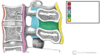
Name the bones of the cranial base

A
  • Frontal
  • Sphenoid
  • Ethmoid
  • Occipital
  • Parietal
  • Temporal
6
Q
A
7
Q

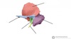
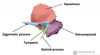
How many face bones are there? Name the face bones

A

14 bones

Zygomatic x2 – cheek bones of the face.
Lacrimal x2
Nasal x2
Inferior nasal conchae x2
Palatine x2 –
Maxilla x2 –
Vomer –
Mandible –

8
Q

What is the TMJ? Where is it located?

A

Articulation between mandible and temporal bone. Located at ends of mandible

9
Q

What is the purpose of the inferior nasal conchae? Where is it located?

A

located within the nasal cavity. Serve to increase SA of the nasal cavity and therefore the amount of inspired air that can come into contact with the cavity walls.

10
Q

What is the purpose of the palatine bone?

A

Forms part of hard palate at rear of oral cavity

11
Q

What does the maxilla help to form?

A

hard palate

12
Q

what does the vomer form?

A

Posterior aspect of the nasal septum

13
Q

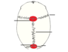
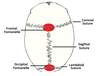
Why are sutures of the skull clinically relevant? Name the 3 most clinically relevant. What are fontanelles? Name them

A

Points of potential weakness.

Coronal, sagittal, lambdoid sutures

Fontanelles (incompletely fused suture joints in neonates). Frontal fontanelle and occipital fontanelle

14
Q
A
15
Q

What is the pterion? Where is it located? What happens if this were to be fractured?

A

Thinnest part of the skull. Fracture here can lacerate an underlying artery (middle meningeal artery) and result in an extradural haematoma (blood collects in between dura mater and skull, increasing intracranial pressure). Symptoms are nausea, vomiting, seizures, bradycardia.

16
Q

How do you treat an extradural haematoma?

A

Treated with diuretic in minor cases and drilling burr holes into skull in more extreme cases

17
Q

What is the anterior cranial fossa?

A

Depression of skull formed by frontal, ethmoid and sphenoid bones

18
Q

What is the middle cranial fossa?

A

Depression formed by sphenoid, temporal and parietal bones

19
Q

What is the posterior cranial fossa?

A

Depression formed by squamous and mastoid temporal bone, and occipital bone

20
Q

What are the 4 types of cranial fractures? How does basal skull fracture present?

A

Depressed – Bone depresses inwards. Possible brain injury,
Linear – Break in bone traversing its full thickness. Most common.
Basal Skull – Affects base of the skull. Presents with bruising behind ears, known as ‘Battle’s sign’ or bruising around the eyes / orbits, known as Raccoon eye’s.
Diastatic – Fracture that occurs along a suture line causing a widening of the suture. Most often seen in children.

21
Q
A
22
Q

What is the function of the squamous part of the temporal bone?

A

Lower part is the site of origin of the temporalis muscle

23
Q

What is the function of the zygomatic process of the temporal bone?

A
  • Articulates with the zygomatic bone, forming the zygomatic arch (cheek bones)
  • Masseter muscles attaches some fibres to lateral surface of the zygomatic process.
  • Articular tubercle on the zygomatic arch forms part of the temporomandibular joint.
24
Q

What is the function of the tympanic part of the temporal bone?

A

Surrounds the external auditory opening

25
Q

What is the function of the petrous part and the mastoid part of the temporal bone?

A
  • Mastoid process, inferior projection of the bone, is palpable just behind the ear and is a site of attachment for SCM.
  • Mastoid air cells – Hollowed out areas within the temporal bone that act as a reservoir of air, equalising the pressure within the middle ear in cases of auditory tube dysfunction. Can become infected, known as mastoiditis.
  • Petrous part lies at base of temporal bone and contains inner ear
26
Q

What is mastoiditis? Where can it spread to?

A

Infection of mastoid process can spread into the middle cranial fossa and into the brain causing meningitis.

27
Q

What is the function of the styloid process?

A

Acts as an attachment point for muscles and ligaments

28
Q

What are the features of cervical vertebrae?

A
  • Triangular vertebral foramen
  • Bifid spinous processes
  • Transverse foramina
29
Q

What are the features of the atlas? What allows it to articulate with the dens?

A
  • No vertebral body
  • No spinous processes
  • Anterior articular facet which articulates with the dens of the axis
  • Posterior arch has a groove for the vertebral artery and C1 spinal nerve.
30
Q

What are the 2 parts to the atlanto-axial joint?

A
  • Has a medial and 2 lateral aspects
  • Medially – Made up of dens process and atlas facet for dens. Pivot type synovial joint.
  • Laterally – Made up of articulation between the inferior facets of C1 and the superior facets of C2. Plane type synovial joints.
31
Q

What is the atlanto-occipital joint?

A
  • Articulation between the spine and cranium
  • Occurs between the superior facets of the Atlas and the occipital condyles at the base of the cranium.
  • Condyloid type synovial joints
32
Q

What are vertebral arch joints?

A

Formed by articulation of the superior and inferior articular processes of adjacent vertebrae

33
Q

What ligaments are found throughout the spine? What is found only in the cervical spine?

A

Throughout:

  • Interspinous ligament
  • Anterior and posterior longitudinal ligaments
  • Ligamentum flavum

Only in cervical spine:

  • Nuchal ligament
  • Transverse ligament of Atlas
34
Q
A
35
Q

What is the function of the nuchal ligament?

A

Continuation of supraspinous ligament. Attaches tips of the spinous processes from C1-C7. Provides proximal attachment for the rhomboids and the trapezius.

36
Q

What is the function of the transverse ligament of Atlas?

A

Connects the lateral masses of the Atlas, and anchors the dens in place

37
Q

Through which transverse foramina does the vertebral artery travel?

A

C2-6

38
Q

How many cervical spinal nerves are there? Explain

A
  • At C7, there are spinal nerves extending from above and below this vertebra
  • There are 8 spinal nerves associated with 7 cervical vertebrae.
39
Q

Where does the vertebral artery run at C1?

A

Vertebral artery runs along the groove for the vertebral artery.

40
Q

What is a jefferson fracture of the atlas? How does it occur? What happens?

A
  • Occurs due to a vertical fall onto an extended neck
  • Results in compression of the lateral masses of the Atlas, driving them apart and fracturing one or both of the posterior / anterior arches.
  • Can also rupture the transverse ligament of the Atlas.
41
Q

What occurs in a hyperxtension / whiplash injury? Minor case, severe case, and worst case scenarios?

A
  • Minor cases result in damage to the anterior longitudinal ligament of the spine
  • More severe cases fractures to any cervical vertebrae can occur due to sudden compression by rapid deceleration.
  • Worse case scenario is the dislocation or subluxation of the cervical vertebrae. Often happens at C2 level. Can lead to spinal cord injury, quadraplegia or death.
42
Q

What is a hangmans’s fracture? What results?

A
  • Fracture of the pars interarticularis, a bony column between the superior and inferior articular facets of the axis.
  • Likely to rupture the spinal cord and result in death.
43
Q

What is a den’s fracture? What is it commonly caused by and what results?

A
  • Commonly caused by falls and traffic collision
  • High risk of avascular necrosis
  • Slight risk of spinal cord involvement.
44
Q
A