Misc Flashcards

1
Q
A

COPS - 3977

Ciliary (3), Otic (9), Pterygopalatine (7), Submandibular (7)

2
Q

What is a rolling and a sliding hernia?

A

Rolling - Gastroesophageal junction in place and gastric fundus herniates upwards

Sliding - Cardia of stomach herniates upwards into oesophagus

3
Q

Where can an infection spread between the investing and visceral part of the pretracheal fascia?

A

inferiorly into the chest, causing infection of the anterior mediastinum.

4
Q

Where can an infection Posterior to the prevertebral fascia spread?

A

Retropharyngeal space, down into mediastinal contents

5
Q
A
6
Q

At what spinal level do oyu palpate the carotid?

A

C6

7
Q

What spinal level does the carotid bifurcate? What about trachea and aorta?

A

Carotid - C4

Trachea - T4

Aorta - L4

8
Q

What nerve controls the dilator pupillae?

A

Sympathetic nervous system

9
Q

What nerve controls sphincter pupillae?

A

Ciliary ganglion

10
Q

What is the most common non trauma cause of facial paralysis? Where does it occur? What happens?

A
  • Most common non trauma cause is inflammation of the facial nerve near its exit from the cranium at the stylomastoid foramen:
    • Inflammation causes edema which compresses the nerve at the intracranial facial canal.
    • Results in affected area sagging
11
Q

What structure does the facial nerve pass through? Why is this clinically relevant? Where can disease of this structure refer to?

A
  • Nerve passes through the parotid gland and therefore vulnerable to injury during surgery on the gland or disease of the gland:
    • Parotid gland disease can cause pain in the temporal region and auricle of the ear.
12
Q
A
13
Q
A
14
Q

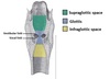
What are the green, orange, purple and red fascia in the pic?

A

Green - investing layer

Purple - Pretracheal layer

Orange - Prevertebral layer

Red - Carotid sheath

15
Q

What is the platysma? Where is it found and what supplies it? What is its function?

A
  • Broad thin sheet of muscle in the superficial cervical fascia.
  • Supplied by the facial nerve
  • Depresses the mandible and draws the corners of the mouth inferiorly.
16
Q

Where can an infection spread:

a) between investing and muscular pretracheal
b) between investing and visceral pretracheal
c) between prevertebral and investing

A

a) cant spread beyond manubrium (superior part of sternum)
b) can spread into thoracic cavity anterior to pericardium
c) can spread laterally in the neck, may perforate fascial layer and enter retropharyngeal space resulting in a bulge in the pharynx and difficulty swallowing and speaking (dysphasia and dysphonia).

17
Q

What is the borders of the anterior triangle of the neck?

A

Superiorly – Inferior border of the mandible (jawbone)
Laterally – Medial border of the sternocleidomastoid
Medially – Imaginary sagittal line down midline of body

18
Q

What are the borders of the posterior triangle of the neck?

A

Anterior: Posterior border of the SCM.
Posterior: Anterior border of the trapezius muscle.
Inferior: Middle 1/3 of the clavicle.

19
Q
A
20
Q
A
21
Q
A
22
Q

What are the 4 types of cranial fractures? How does basal skull fracture present?

A

Depressed – Bone depresses inwards. Possible brain injury,
Linear – Break in bone traversing its full thickness. Most common.
Basal Skull – Affects base of the skull. Presents with bruising behind ears, known as ‘Battle’s sign’ or bruising around the eyes / orbits, known as Raccoon eye’s.
Diastatic – Fracture that occurs along a suture line causing a widening of the suture. Most often seen in children.

23
Q
A
24
Q

What are the features of cervical vertebrae?

A
  • Triangular vertebral foramen
  • Bifid spinous processes
  • Transverse foramina
25
Q

Through which transverse foramina does the vertebral artery travel?

A

C1-6

26
Q

What occurs in a hyperxtension / whiplash injury? Minor case, severe case, and worst case scenarios?

A
  • Minor cases result in damage to the anterior longitudinal ligament of the spine
  • More severe cases fractures to any cervical vertebrae can occur due to sudden compression by rapid deceleration.
  • Worse case scenario is the dislocation or subluxation of the cervical vertebrae. Often happens at C2 level. Can lead to spinal cord injury, quadraplegia or death.
27
Q
A
28
Q

Why can injuries to scalp cause excessive bleeding?

A
  1. Walls of arteries bound tightly to underlying connective tissue of scalp, preventing constriction to limit blood loss.
  2. Numerous anastomoses formed by arteries which produce a densely vascularised area
29
Q

What is the middle meningeal artery a branch of?

A

Maxillary artery

30
Q

Through which transverse processes does the vertebral artery travel through?

A

C6 - 1

31
Q

Label the branches of the thyrocervical trunk

A
32
Q

What are the venous drainages of:

a) brain and meninges
b) scalp and face
c) neck

A

Brain and meninges – Dural venous sinuses
Scalp and face – Veins synonymous with arteries of the face and scalp. Drain into internal and external jugular veins
Neck – Anterior jugular veins

33
Q

How does the IJV exit the skull?

A

Jugular foramen

34
Q

Where is the cavernous sinus located?

A

Lateral aspect of sphenoid bone

35
Q

What nerves are located in the lateral wall of the cavernous sinus?

A

the oculomotor (III), trochlear (IV), ophthalmic (V1) and maxillary (V2) nerves

36
Q
A
37
Q

Which nodes are paired and unpaired?

A
  • Paired - palatine tonsil, tubal tonsil
  • Unpaired – pharyngeal (adenoid) tonsil, lingual tonsil
38
Q

Where does pus accumulate in a retropharyngeal abscess? What happens in a retropharyngeal abscess?

A

In a retropharyngeal abscess, puss accumulates in space between prevertebral fascia and buccopharyngeal membrane. Can result in compression of pharynx à dysphagia and dysarthria (difficulty speaking).

39
Q

What are the 3 extra classes of special cranial nerves? What do they do?

A
  1. Special visceral efferents – muscles derived from pharyngeal arches (CNV, VII, IX, X)
  2. Special somatic afferents – equilibration, hearing, and sight
  3. Special visceral afferents – taste
40
Q

What are the names of the 12 cranial nerves?

A

I - Olfactory

II - Optic

III - Oculomotor

IV - Trochlear

V - Trigeminal

VI - Abducent (Abducens)

VII - Facial

VIII - Vestibulocochlear

IX - Glossopharyngeal

X - Vagus

XI - Spinal Accessory

XII - Hypoglossal

Oh Oh Oh To Touch And Feel Very Girly Vaginas So Heavenly

41
Q

What is the function of the 12 cranial cranial nerves?

A

I - Sensory

II - Sensory

III - Motor

IV - Motor

V - Both

VI - Motor

VII - Both

VIII - Sensory

IX - Both

X - Both

XI - Motor

XII - Motor

Some Say Marry Money But My Brother Says Big Boobs Matter More

42
Q

What are the 3 cervical sympathetic ganglia? What are they known as after synapsing? Where do they travel?

A

Superior, middle and inferior

After synapsing, known as internal and external carotid nerves. Travel alongside BVs to target tissues.

43
Q

Where does the ciliary ganglion lie?

A

Orbital cavity

44
Q

What causes Horner’s Syndrome? How does it manifest?

A

Interruption of a cervical sympathetic trunk

Features on ipsilateral side of head:

  • Miosis - constriction of pupil
  • Ptosis
  • Vasodilation - Redness and increased temp of skin
  • Anhydrosis - Absence of sweating.
45
Q
A
46
Q

What muscles are reponsible for protrusion and retraction of the mandible?

A

Lateral pterygoid muscle – protrusion
temporalis and digastric muscle – retraction

47
Q

Why is an infection of the pterygoid region a danger to the eye?

A

pterygoid venous plexus connects to opthalmic and anterior facial veins. Pus inside pterygoid venous plexus raises pressure inside opthalmic veins or lead to cavernous sinus thrombosis, resulting in paralysis of extraocular muscles

48
Q

What is an auricular haematoma? What does it result from? What can it result in?

A

Blood collects between cartilage and pericondrium overlying it
Usually results from trauma
Accumulation of blood disrupts vascular supply to cartilage of the pinna. If it is not drained, can result in cauliflower ear.

49
Q

In which bone does the middle ear lie?

A

temporal

50
Q
A
51
Q

What are the roof and floor borders of the middle ear

A

Roof – Petrous part of temporal bone
Floor – Jugular wall

52
Q

What are the lateral and medial borders of the middle ear? What nerve travels in the medial wall?

A

Lateral wall – TM
Medial wall – Internal ear wall. Has bulge due to facial nerve which travels nearby

53
Q

What are the anterior and posterior borders of the middle ear? What structures go through the anterior wall? What is the hole in the posteriro wall called?

A

Anterior wall – thin bony plate with openings for auditory tube and tensor tympani muscle
Posterior wall – AKA mastoid wall. Bony partition between Epitympanic recess and mastoid air cells. Hole in this partition called the aditus to the mastoid antrum.

54
Q

What is a cholesteastoma? How does it cause damage and how does it present?

A
  • This is a growth of stratified squamous epithelium in the middle ear
  • Can be congenital or acquired
  • Grows and causes damage to bones of middle ear by increased pressure or release of osteolytic enzymes
  • Patients present with hearing loss and may have facial nerve palsy due to close proximity of facial canal. Treat surgically.
55
Q

What innervates the 2 middle ear muscles? Which bones do they attach to?

A

Tensor tympani attaches to malleus, innervated by mandibular

Stapedius attaches to stapes and innervated by facial

56
Q

What is glue ear?? Why does it occur? What is the result and how does it present?

A
  • Otitis media with effusion
  • Occurs due to persistent dysfunction of auditory tube. If auditory tube unable to equalise middle ear pressure (can be due to blockage, inflammation or genetic mutation), negative pressure develops in middle ear
  • Negative pressure draws out transudate from mucosa of middle ear, creating an environment suitable for pathogens.
  • Inspection of patient will reveal inverted eardrum with fluid visible inside the ear
57
Q

What are the ducts of membranous labyrinth filled with?

A

filled with endolymph

58
Q

How is the cochlear duct held in place?

A

spiral lamina

59
Q

What are the saccule and utricle? What is their function?

A

Membranous sacs in vestibule

Provides information about head position when not moving

60
Q

What is menieres disease? How is it caused?

A
  • Disorder of inner ear – episodes of vertigo, tinnitus and hearing loss
  • Caused by excess accumulation of endolymph, causing progressive distension of the ducts. Resulting pressure fluctuations damages the thin membranes of the ear that detect balance and sound.
61
Q

How does swallowing allow equalisation of pressure?

A

opens up eustachian tube allowing equalisation of pressure from middle ear to outside

62
Q

What are the 4 pathways for air to flow in the nasal cavity?

A

Inferior meatus – between inferior concha and floor of nasal cavity
Middle meatus – lies between inferior and middle conchae
Superior meatus – lies between middle and superior conchae
Spheno-ethmoidal recess – superior and posterior to superior concha

63
Q
A

SENSE

64
Q

What structures pass through the sphenopalatine foramen?

A

Sphenopalatine artery, vein and nerve nerves pass through here.

65
Q

What internal carotid branches supply the nose? How do they reach the nasal cavity?

A

Anterior and posterior ethmoidal artery
Descend into nasal cavity through cribiform plate

66
Q

What external carotid branches supply the nose?

A

Sphenopalatine artery
Greater palatine artery

67
Q

Where do the veins drain from the nasal cavity in only some individuals and why is this clinically relevant?

A

In some individuals, some nasal veins join with sagittal sinus, a potential pathway from infection to spread from nose into cranial cavity.

68
Q
A
69
Q

Describe the general innervation of the septum, lateral walls, and skin of nose

A

Septum and lateral walls innervated by nasopalatine nerve and nasociliary nerve
Skin of nose innervated by trigeminal nerve

70
Q

Where is the trigeminal ganglion found?

A

middle cranial fossa

Lateral to cavernous sinus in a depression in temporal bone known as trigeminal cave

71
Q

Through which foramen do the ophthalmic and maxillary nerves travel?

A

Ophthalmic - superior orbital fissure

Maxillary - foramen rotundum

72
Q

Through which foramen does the mandibular nerve travel through?

A

foramen ovale

73
Q

What is the corneal reflex? What nerves are involved and what is an absent reflex indicative of?

A
  • Involuntary blinking of eyelids stimulated by stimulation of cornea
  • Ophthalmic nerve acts as afferent limb, facial nerve is the efferent limb.
  • If reflex absent à sign of damage to the trigeminal/ophthalmic nerve or facial nerve.
74
Q

What nerves does the mandibular nerve give rise to?

A

Gives rise to buccal, inferior alveolar, auricotemporal, and lingual nerve

75
Q

Where does the inferior alveolar nerve travel through?

A

Mandibular foramen and mandibular canal.

76
Q

Why is the inferior alveolar nerve blocked for some dental procedures? Where is it blocked? Where is anaesthetic administered?

A
  • Inferior alveolar nerve blocked for some dental procedure, before it gives rise to plexus
  • Anaesthetic administered at mandibular fossa.
77
Q

What is the action of the inferior rectus, medial rectus and inferior oblique?

A

Inferior rectus – depresses eyeball
Medial rectus – adducts eyeball
Inferior oblique – Elevates, abducts, and laterally rotates eyeball

78
Q

What are 3 anatomical causes of an oculomotor nerve lesion?

A

Increases intracranial pressure – compresses nerve against temporal bone
Aneurysm of posterior cerebral artery
Cavernous sinus infection or trauma

79
Q

What is the result of an oculomotor nerve lesion?

A

Ptosis due to paralysis of LPS
Eyeball resting in down and out position due to paralysis of superior, inferior, medial rectus
Dilated pupil due to unopposed action of dilator pupillae muscle

80
Q

Why can a tumour of the middle ear cause abnormal sensation in taste?

A

compression of facial nerve by tumour can damage chorda tympani branch that supplies special taste sensation to anterior 2/3rds of tongue

81
Q

Why is the inferior thyroid artery tortuous?

A

thyroid gland ascends when swallowing. Tortuous to allow the artery to ascend with thyroid gland

82
Q

What is the function of the carotid body and carotid sinus? Where are they located?

A

Carotid body - chemoreceptor, oxygen levels, part of artery

Carotid sinus - baroreceptor, pressure levels, outside artery

83
Q

What nerves can be damaged in the neck?

A

vagus and sympathetic cervical chain

84
Q

Why might an infection lead to blockage of the eustachian tube? What disease does this lead to?

A

Lymphoid tissue forms a Tonsillar ring around the superior part of the pharynx, which aggregates to form the Adenoids. The adenoids may become swollen during infection in children

Leads to otitis media

85
Q
A
86
Q

What are the 3 muscles of the inner longitudinal muscle layer? What is their function?

A
  • Stylopharyngeus
  • Palatopharyngeus
  • Salpingopharyngeus

Act to shorten and widen pharynx

87
Q

What is the sensory innervation of the pharynx?

A

Glossopharyngeal nerve

Nasopharynx is via opthalmic and maxillary divisions of trigeminal

88
Q

What is the motor innervation of the pharynx?

A

Vagus nerve

Except stylopharyngeus muscle which is glossopharyngeal nerve

89
Q

What are the arteries that supply the pharynx? Which artery do they originate from?

A

From external carotid:

  • Ascending pharyngeal
  • lingual
  • facial
  • maxillary
90
Q

What is quinsy? How is it treated?

A

Peritonsillar abscess pushing uvula to other side

Requires drainage

91
Q

Name the 3 regions food is likely to become stuck in the oropharynx

A
  • Vallecula - pouch between tongue and epiglottis
  • Base of tongue
  • Region of palatine tonsil
92
Q
A
93
Q

What is the nervous supply of the intrinsic muscles of the larynx?

A

The Recurrent Laryngeal Nerve supplies the intrinsic muscles. The exception is the Cricothyroid Muscle, which is supplied by the External Laryngeal Nerve.

94
Q

Which muscles are responsible for the abduction and adduction og the vocal cords?

A

Abduction – Posterior Cricoarytenoid

Adduction – Lateral Cricoarytenoid and cricothyroid

95
Q

What is the afferent and efferent limb of the gag reflex?

A

Afferent - glossopharyngeal

Efferent - Vagus

96
Q

What is a pharyngeal pouch?

A

Weakening between cricopharyngeus and thyropharyngeus muscles leading to diverticulum of pharyngeal mucosa between them

97
Q

What is the precursor of the anterior lobe of the pit gland and its origin?

A

Precursor - Rathke’s pouch

Origin - ectoderm

98
Q

What is the origin of posterior lobe of pit gland?

A

Origin - neuroectoderm

99
Q

Describe how the pituitary gland is formed.

A
  1. Infundibulum extends down towards roof of oral cavity
  2. Outpushing from roof grows up to meet it (Rathke’s Pouch) and it oses its connection with roof of mouth
  3. Pouch comes to lie anterior to infundibulum
  4. Pouch differentiates to cells of ant pit.
  5. Hypophyseal portal system links hypothalamus to pituitary gland.
100
Q

Whats the name of the 3 medial lingual swellings and which PA is each derived from?

A

1st PA - Tuberculum impar

2nd and 3rd PA - cupola

4th PA - Epiglottal swelling

101
Q

Describe the development of the tongue

A
  1. Lateral swelling overgrow tuberculum impar
  2. 3rd Arch part of cupola overgrows its second Arch component
  3. Degeneration occurs, freeing tongue from floor of oral cavity except lingual frenulum
102
Q

What innervates the special sensation of tongue?

A

anterior 2/3rds - Chorda Tympani

posterior 1/3rd - Glossopharyngeal

103
Q

What is the general innervation of the tongue?

A

Anterior 2/3rds - Lingual (comes from mand branch of trigeminal)

Posterior 1/3rd - Glossopharyngeal

104
Q

What innervates the muscles of the tongue?

A

All muscles except palatoglosssus - Hypoglossus nerve

Palatoglosssus - Vagus nerve

105
Q

Where does the thyroid gland first appear in development?

A

Between tuberculum Impar and cuploa of tongue at site called foramen cecum

106
Q

What is first arch syndrome? What is the usual result

A

failure of colonisation of 1st arch with neural crest cells

Usual result is hypoplasia of mandible and facial bones

107
Q

What is CHARGE syndrome? What is it caused by?

A

CHARGE

Coloboma (hole in iris
Heart defects
Atresia (blockage of posterior nasal cavity)
Retardation of growth and development
Genital hypoplasia
Ear defects

CHD7 mutation, essential for production of multipotent nerual crest cells

108
Q

State the borders of the bony orbit - Roof, medial, lateral, floor, apex.

A

Roof - frontal

Medial - lacrimal, ethmoid, maxilla

Lateral - zygomatic

Floor - Maxilla, zygomatic

Apex - Sphenoid

109
Q

What structures run through the superior orbital fissure?

A

Large French Tarts Sit Nakedly In Anticipation of Sweets

Lacrimal nerve, frontal nerve, trochlear nerve, sup oculomotor, nasociliary nerve, inf oculomotor, abducens, opthalmic veins (sup), sympathetic nerves

110
Q

What structures run through the inferior orbital fissure?

A

Maxillary nerve, inferior opthalmic veins, sympathetic nerves

111
Q

What is an orbital rim fracture? What results?

A

Fracture of lateral bones. Likely to occur at sutures

rsults in exophthalmos, increase in intraorbital pressure, and possibly haemorrhage

112
Q

What is a blowout fracture of bony orbit?

A

Fracture of Medial and inferior walls. Herniating into ethmoid and maxillary sinuses

113
Q

What nerve innervates the LPS and the tarsal muscle?

A

LPS - oculomotor

Tarsal - sympathetic

114
Q

How do the eye muscles move?> What is their innervation?

A

LR6 SO4 R3

115
Q

What is the symptoms of a trochlear nerve lesion?

A

Diplopia and a head tilt away from site of lesion

116
Q

What is the resting position of an abducens nerve lesion?

A

Eye adducted by MR

117
Q

What is the effect on the eye of a facial nerve palsy?

A

Inability to close eyes due to paralysed orbicularis oculi

Loss of blink and corneal reflex

Tears pool in eyelids and can cause infection

118
Q

What nerve is responisble for production of tearS?

A

ps fibres of facial

119
Q

What is a stye and a meiobomian cyst?

A

Stye - infection of gland at base of eyelash

Meiobomian cyst - blocked tarsal gland

120
Q
A