8 Ventilation and Perfusion Flashcards
(28 cards)
Ventilation
- The repetitive movement of gas into and out of the lungs
- Delivers the oxygen and removes the carbon dioxide that is exchanged across the alveolar-capillary interface

Lung volumes and capacities
- Tidal volume (VT)
- Residual volume (RV)
- Expiratory reserve volume (ERV)
- Inspiratory reserve volume (IRV)
-
Tidal volume (VT)
- Volume of gas inhaled (or exhaled) during a breath
- ~500ml in a resting adult
- Increases as needed to meet the metabolic demands of the body (e.g. during exercise)
-
Residual volume (RV)
- Amount of gas remaining in the lungs after a maximal expiration
- Determined primarily by the inward pressure generated by the expiratory muscles and by the outward elastic recoil of the respiratory system
- ~1.5L in a normal adult
-
Expiratory reserve volume (ERV)
- Volume of gas that can be forced from the lungs starting at the end of a normal tidal expiration
-
Inspiratory reserve volume (IRV)
- Volume of gas that can be inhaled during a maximal inspiration starting at the end of a normal tidal inspiration

Lung volumes and capacities
- Functional residual capacity (FRC)
- Total lung capacity (TLC)
- Vital capacity (VC)
- Inspiratory capacity (IC)
-
Functional residual capacity (FRC)
- Volume remaining in the lungs at the end of a passive expiration
- Represents the equilibrium position of the respiratory system
- The point at which the inward elastic recoil of the lungs is balanced by the outward recoil of the chest wall
- Sum of the residual and the expiratory reserve volumes
- FRC = RV + ERV
-
Total lung capacity (TLC)
- Volume in the lungs at the end of a maximal inspiration
- Determined by the maximum force generated by the inspiratory muscles and by the inward elastic recoil of the lungs and chest wall
-
Vital capacity (VC)
- Volume of gas that can be exhaled during a maximal effort beginning at the end of a maximal inspiration
- Sum of the IRV, ERV, and VT
- Difference between TLC and RV
- VC = TLC - RV
-
Inspiratory capacity (IC)
- Amount of gas that enters the lungs during a maximal inspiration beginning at the end of a normal tidal expiration
- Sum of the IRV and VT
- IC = IRV + VT

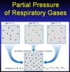
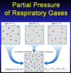
Partial pressure of respiratory gases
- In a gas mixture, the pressure exerted by a gas is equal to…
- Dry air composition of O2, N2, & CO2
- Total barometric pressure (PB) at sea level
- Partial pressures of O2, N2, & CO2
-
In a gas mixture, the pressure exerted by a gas is equal to…
- The product of its fractional concentration (F) and the total pressure of all the gases in the mixture
- Pgas = Ptotal x Fgas
-
Dry air composition of O2, N2, & CO2
- FO2 = 0.21 (21%)
- FN2 = 0.79 (79%)
- FCO2 = 0.0004 (0.04%)
-
Total barometric pressure (PB) at sea level
- 760 mmHg
-
Partial pressures of O2, N2, & CO2
- PO2 = PB x F?
- PO2 = 760 x 0.21 = 160 mmHg
- PN2 = 760 x 0.79 = 600 mmHg
- PCO2 = 760 x 0.0004 = 0.3 mmHg

Partial pressure of respiratory gases
- Partial pressure of the water vapor (PH2O)
- When air enters the conducting airways of the lungs…
- Calculation of partial pressure of inspired gas in the airways (PIgas)
- Partial pressures of O2, N2, & CO2
-
Partial pressure of the water vapor (PH2O)
- ~47 mmHg at body temperature
- When air enters the conducting airways of the lungs…
- It is heated and humidified
- Total pressure exerted by dry gas decreases
- Partial pressure of each gas falls
- Calculation of partial pressure of inspired gas in the airways (PIgas)
- PIgas = (PB – PH2O) x Fgas
-
Partial pressures of O2, N2, & CO2
- PIO2 = (760 – 47) x 0.21 = 150mmHg
- PIN2 = (760 – 47) x 0.79 = 150mmHg
- PICO2 = (760 – 47) x 0.0004 = 150mmHg

Partial pressure of respiratory gases
- Once gas enters the alveoli…
- The partial pressure of gases in the alveoli…
- For practical purposes, we can assume that the mixed alveolar PCO2 (PACO2)…
- Once gas enters the alveoli…
- Diffusion occurs leading to a drop in the PO2 and an increase in PCO2
- The partial pressure of gases in the alveoli…
- Cannot be measured, but they can be estimated
- For practical purposes, we can assume that the mixed alveolar PCO2 (PACO2)…
- Is equal to the PCO2 of arterial blood (PaCO2)

Partial pressure of respiratory gases:
Alveolar air equation
- PAO2
- PA-aO2 gradient
-
Used to calculate the average alveolar PO2 (PAO2) assuming “ideal” conditions (i.e. no mismatching of ventilation and perfusion)
- PAO2 = [(PB – PH2O) * FIO2] – [PACO2 / R]
- PACO2 = PaCO2 = 40 mmHg
- R = VCO2 / VO2 = 0.8
- PAO2 = [(760 - 47) * 0.21] - [40 / 0.8] = 100 mmHg
-
Used to derive the PA-aO2 or the A-a gradient
- Difference between the calculated alveolar and the measured arterial PO2 (always exists)
- Normally 8-12 mmHg
- Increases in the presence of ventilation-perfusion mismatching, shunt, and diffusion impairment

Partial pressure of respiratory gases
- Respiratory exchange ratio (R)
- Partial pressure of O2, CO2, & N2 in the alveoli
- Respiratory exchange ratio (R)
- Volume of CO2 that enters the alveoli divided by the volume of O2 that diffuses into the pulmonary capillary blood over a given period of time (VCO2/VO2)
- Ratio of CO2 produced to O2 consumed by the tissues
- Assumed to be 0.8
- There is more O2 leaving the lung than CO2 entering it
- PO2 will fall by more than the increase in PACO2
- It will decrease by PACO2/R
- Volume of CO2 that enters the alveoli divided by the volume of O2 that diffuses into the pulmonary capillary blood over a given period of time (VCO2/VO2)
-
Partial pressure of O2, CO2, & N2 in the alveoli
- PAO2 = 100 mmHg
- PACO2 = 40 mmHg
- PAN2 = 573 mmHG

The oxygen cascade
- Wordy explanation
- Numeral explanation
- Wordy explanation
- Air comes in at 160
- Once it enters our conducting airways, it’s 150
- Then it drops to 100 by the time it gets to the alveoli
- Difference b/n alveolar gas and arteriolar blood: A-a gradient
- As the blood is carried to the tissues, the PO2 really drops
- By the time it gets to the capillaries, there’s very little PO2
- Estimated mitochondria PO2 = 5
- Numeral explanation
- Air
- PO2 = 160
- PCO2 = 0
- Airways
- PO2 = 150
- PCO2 = 0
- Alveoli
- PO2 = 100
- PCO2 = 40
- Arterial
- PO2 = 95
- PCO2 = 40
- Mixed venous
- PO2 = 40
- PCO2 = 46
- Air

Dead space
- Dead space
- Alveolar dead space
- Physiologic dead space
-
Dead space
- A significant portion of each tidal breath never reaches the alveoli
- Volume of gas that fills the nose, mouth, pharynx, larynx, and conducting airways
- Since this gas is never in contact with pulmonary capillary blood, it does not participate in gas exchange
- Varies with body (and airway) size
- Although it can be measured, for practical purposes it is often estimated as 1ml per pound of ideal body weight
- Alveolar dead space
- Another portion of the tidal breath that reaches alveoli that either
- Receive no blood flow
- Are under-perfused relative to the amount of ventilation they receive
- Another portion of the tidal breath that reaches alveoli that either
- Physiologic dead space
- Sum of the anatomic and alveolar dead space
Volumes (L)
- Tidal volume (VT)
- Dead space
- Dead space volume (VD)
- Alveolar volume
- Tidal volume (VT)
- Volume of gas that enters the lungs during a single breath
-
Dead space
- A significant portion of each tidal breath never reaches the alveoli
- Volume of gas that fills the nose, mouth, pharynx, larynx, and conducting airways
- Since this gas is never in contact with pulmonary capillary blood, it does not participate in gas exchange
- Varies with body (and airway) size
- Although it can be measured, for practical purposes it is often estimated as 1ml per pound of ideal body weight
-
Dead space volume (VD)
- Amount of gas entering the physiologic dead space
- The volume of each breath that does not participate in gas exchange
- Anatomic + alveolar dead space
-
Alveolar volume (VA)
- Volume of gas that actually reaches the alveoli and participates in gas exchange during a tidal breath
-
Difference between tidal volume and dead space volume
- VA = VT - VD
- If take a breath in of 500 ml and have a dead space of 200 ml, then true alveolar volume that participates in gas exchange is 300 ml

Ventilations (L/min)
- Minute ventilation (VE)
- Dead space ventilation (VD)
- Alveolar ventilation (VA)
-
Minute ventilation (VE)
- Total volume of gas that enters or leaves the lungs each minute
- Product of tidal volume and respiratory rate (RR)
- VE = VT x RR
-
Dead space ventilation (VD)
- Volume of gas that enters and leaves the physiologic dead space each minute
- VD = VD x RR
-
Alveolar ventilation (VA)
-
Volume of gas entering and leaving the lungs each minute that participates in gas exchange
- VA = VA x RR
- Difference between minute ventilation and dead space ventilation
- VA = VE – VD
-
Volume of gas entering and leaving the lungs each minute that participates in gas exchange
The influence of alveolar ventilation on PACO2
- The partial pressure of carbon dioxide in the alveolar gas (PACO2) is determined by…
- CO2 removal from alveoli vs. alveolar ventilation
- PACO2 vs. VA
- PACO2 equations
- PaCO2 equations
-
PACO2 and PaCO2 are directly related to…
- The rate at which CO2 enters the alveoli
-
The partial pressure of carbon dioxide in the alveolar gas (PACO2) is determined by…
- The rate at which CO2 is produced by the tissues (VCO2)
- The rates at which CO2 enters and leaves the alveoli
- –> proportional to the partial pressure of CO2 in mixed venous blood (PvCO2)
- –> –> depends on the rate of CO2 production by the tissues (VCO2)
- PACO2 α VCO2
- CO2 removal from alveoli vs. alveolar ventilation
- The rate at which CO2 is removed from the alveoli is directly proportional to alveolar ventilation
-
PACO2and PaCO2 vs. VA
-
PACO2 and PaCO2 are inversely related to the rate at which CO2 is removed from the alveoli
- Determined by alveolar ventilation (VA)
- PACO2 α 1 / VA
-
PACO2 and PaCO2 are inversely related to the rate at which CO2 is removed from the alveoli
- PACO2 equations
- PACO2 α VCO2 / VA
- PACO2 = K x VCO2 / VA
- PaCO2 equations
- Alveolar and arterial PCO2 are essentially equal
- PaCO2 = K x VCO2 / VA
- PaCO2 = K x VCO2 / (VE - VD)
-
PaCO2 = K x VCO2 / VE [1 – (VD / VT)]
- As VD/VT rises, VE must increase to maintain the same PaCO2

The influence of alveolar ventilation on PAO2
- Unlike PACO2 (and PaCO2), alveolar ventilation does not directly affect PAO2
- Instead, its effect is indirect and produced by changes in PACO2
- By examining the alveolar air equation, it is evident that PAO2 and PACO2 must move in opposite directions
- When PACO2 increases, PAO2 decreases
- When PACO2 decreases, PAO2 increases
Regional distribution of alveolar ventilation
- In normal subjects, who are either seated or standing, the volume of air reaching the alveoli…
- Although a single value is often used to describe the pressure in the pleural space, in an upright subject, pleural pressure…
- Since the pressure in all alveoli is zero (atmospheric) at the end of a passive exhalation, the transpulmonary pressure…
- During inspiration, trans-pulmonary pressure…
- Since alveoli in the non-dependent regions already have a relatively high volume, they are…
- In normal subjects, who are either seated or standing, the volume of air reaching the alveoli…
- Increases from the apex to the base of the lungs
- This causes ventilation to increase from the top to the bottom of the lungs
- This can be explained by gravity-induced changes in pleural pressure
- Although a single value is often used to describe the pressure in the pleural space, in an upright subject, pleural pressure…
- Progressively increases (becomes less negative) between the top (apex) and bottom (base) of the lungs
- Since the pressure in all alveoli is zero (atmospheric) at the end of a passive exhalation, the transpulmonary pressure…
- Must be higher in the upper lung zones than in the lower zones
- This means that at end-expiration, alveolar volume is high at the lung apex and progressively falls in the more dependent regions of the lungs
- During inspiration, trans-pulmonary pressure…
- Increases equally regardless of alveolar location
- Since alveoli in the non-dependent regions already have a relatively high volume, they are…
- Less compliant than alveoli in the lower lung zones
- This means that for the same change in trans-pulmonary pressure, the amount of fresh gas entering the alveoli is greater in the dependent than in the nondependent regions of the lungs

Perfusion:
Sources of blood flow to lungs
- Bronchial circulation
- Bronchial arteries
- A large proportion of the deoxygenated bronchial venous blood drains into…
- Pulmonary circulation
- Pulmonary arteries
- As compared with the systemic circulation, the pulmonary arteries have…
-
Bronchial circulation
- Bronchial arteries
- Carry oxygenated blood from the left ventricle
- Originate either directly from the aorta or from intercostal arteries
- Supply oxygen to several structures in the thorax including the conducting airways, the visceral pleura, and the esophagus
- A large proportion of the deoxygenated bronchial venous blood drains into the pulmonary veins, thereby producing an anatomic right to left shunt
- Bronchial arteries
-
Pulmonary circulation
- Pulmonary arteries
- Carry the mixed venous blood from all the tissues of the body
- Provide oxygen to the distal airways and alveoli
-
As compared with the systemic circulation, the pulmonary arteries have…
- Both thinner walls and larger lumens
- Less vascular smooth muscle
- No muscular vessels analogous to systemic arterioles
- Pulmonary arteries
Perfusion:
Important differences between the pulmonary and systemic circulations
- Pulmonary vascular resistance (PVR)
- Pulmonary vascular resistance distribution
- Pulmonary arteries
- Pulmonary vascular resistance (PVR) is normally much less than (approximately one-tenth) that of the systemic circulation
- Pulmonary vascular resistance is fairly evenly distributed between the arteries, capillaries, and veins, whereas arteries and arterioles account for approximately 80% of the systemic vascular resistance
- Pulmonary arteries are much more distensible and compressible than their systemic counterparts
Factors affecting pulmonary vascular resistance:
Active vs. passive factors
- Active factors
- Passive factors
- Active factors
- Pulmonary vascular resistance can be influenced by both neural and humoral factors, which alter vascular smooth muscle tone and vessel caliber
- Passive factors
- The distensibility, compressibility, relative lack of smooth muscle, and low intravascular pressure that characterize the pulmonary circulation cause PVR to be strongly influenced by a variety of extra-vascular or “passive” factors that have no effect on vascular smooth muscle tone
Factors affecting pulmonary vascular resistance:
Active factors:
Neural factors
- The pulmonary circulation is supplied with both sympathetic and parasympathetic innervation
- Parasympathetic stimulation causes vascular dilation and a decrease in PVR
- Increased sympathetic activity leads to vasoconstriction and an increase in PVR
Factors affecting pulmonary vascular resistance:
Active factors:
Humoral factors
- Pulmonary vasoconstriction is caused by…
- Pulmonary vasodilationis caused by…
- As PAO2 falls…
- Alveolar hypoxia
- Hypoxic-induced vasoconstriction
- Pulmonary vasoconstriction is caused by…
- Catecholamines, (e.g. dopamine, norepinephrine, and epinephrine), histamine, PGE2, PGF2α, and thromboxane
- Pulmonary vasodilationis caused by…
- Acetylcholine, PGE1 and PGI2 (prostacyclin)
- As PAO2 falls…
- Vasoconstriction occurs in the pre-capillary vessels
- The mechanism for this increase in vascular tone is unknown, but the effect is not mediated through the autonomic nervous system
- Alveolar hypoxia
- The most important humoral factor
- May have a direct effect on vascular smooth muscle
- May cause the release of other humoral mediators such as histamine, serotonin, or prostaglandins
- Vasoconstriction may occur…
- Locally (e.g. atelectasis)
- Diffusely (e.g. hypoventilation, high altitude)
- Hypoxic-induced vasoconstriction
- Clearly a beneficial response
- Decreases blood flow to regions of the lung that are poorly ventilated
- Unfortunately, this response is relatively weak due to the lack of vascular smooth muscle
Factors affecting pulmonary vascular resistance:
Passive factors:
Lung volume
- Alveolar vessels
- Primarily…
- Affected by…
- During inspiration…
- As lung volume falls below FRC,…
- Extra-alveolar vessels
- Exposed to…
- During a spontaneous inspiration,…
- During forced expiration below FRC,…
- Both the alveolar and extra-alveolar vessels
- A complex relationship exits between lung volume and PVR
-
Alveolar vessels
- Primarily the pulmonary capillaries
- Affected by alveolar volume
-
Resistance varies directly with lung volume
- During inspiration, the increase in alveolar volume causes the capillaries to be compressed and elongated, thereby increasing the resistance of these vessels
- As lung volume falls below FRC, this process is reversed, and PVR decreases
-
Extra-alveolar vessels
- Exposed to pleural pressure
-
During a spontaneous inspiration, pleural pressure falls
- This increases vascular trans-mural pressure and vessel diameter and causes vascular resistance to decrease
- During forced expiration below FRC, pleural pressure rises and resistance increases
- Both the alveolar and extra-alveolar vessels
- Contribute significantly to total resistance
- A complex relationship exits between lung volume and PVR
- PVR is lowest near FRC and progressively rises with both increasing and decreasing lung volume

Factors affecting pulmonary vascular resistance:
Passive factors:
Cardiac output
- In normal subjects, significant increases in cardiac output (e.g. during exercise)…
- Intra-vascular pressure is determined by…
- Increases in cardiac output must be balanced by…
- As flow increases, pressure…
- Both of these processes lead to…
- In normal subjects, significant increases in cardiac output (e.g. during exercise)…
- Have very little effect on pulmonary artery pressure
- Intra-vascular pressure is determined by…
- The product of resistance and flow
-
Increases in cardiac output must be balanced by…
- A fall in PVR and little change in arterial pressure
-
This decrease in vascular resistance is not mediated by alterations in vascular tone, but is instead due to two passive processes
- Recruitment: increase blood flow –> open up vessels that are closed to begin with near the top of the lungs –> reduce resistance
- Distention: distend all other vessels –> increase their radius –> reduce resistance
- Overall: greater CO –> lower pulmonary vascular resistance –> maintain normal pulmonary arterial pressure (even during very active exercise)
- As flow increases, pressure…
- Rises transiently
- Opens or recruits capillaries and other small vessels that had been closed due to insufficient intra-vascular pressure
- This increase in vascular pressure also causes distention of the pulmonary vasculature
- Both of these processes lead to…
- A fall in PVR and minimize any flow-induced increase in pulmonary vascular pressure

Factors affecting pulmonary vascular resistance:
Passive factors:
Gravity
- Intravascular pressure is higher in dependent than in non-dependent lung regions
- The higher the intravascular pressure, the greater the vascular distention and the lower the PVR
- Due to the weight of the liquid, the pressure within a column of water (or blood) is higher at the bottom than near the top
- Similarly, intra-vascular pressure increases in the dependent (bottom) portions of the lungs
- In an upright subject, this pressure gradient causes progressive vascular distention toward the lung bases and narrowing near the apices
The regional distribution of pulmonary blood flow
- Pulmonary blood flow increases in the _ portions of the lungs
- In a seated or standing subject, blood flow…
- In a supine subject, flow increases to the…
- When a subject lies on his or her side, blood flow increases to the…
- Pulmonary blood flow increases in the dependent (bottom) portions of the lungs
- This effect is mediated by gravity-induced increases in intra-vascular pressure, which, in turn, lead to vascular distention, decreased resistance, and increased flow
- In a seated or standing subject, blood flow is greatest at the lung bases (bottom) and least at the apices (top)
- In a supine subject, flow increases to the anatomically dependent (dorsal) portions of the lungs
- When a subject lies on his or her side, blood flow increases to the dependent lung


