Session 1 Flashcards Preview

ESA2 CVS > Session 1 > Flashcards

Flashcards in Session 1 Deck (64)
Loading flashcards...
1
Q

What are the components of the Cardiovascular System?

A
  • Pump - the heart
  • Distribution system - vessels and blood
  • Exchange mechanism - capillaries
  • Flow control - arterioles and pre-capillary sphincters (resistance vessels)
  • Capacitance - veins
2
Q

How does Exchange of substancs between the blood and cells of the body occur?

A

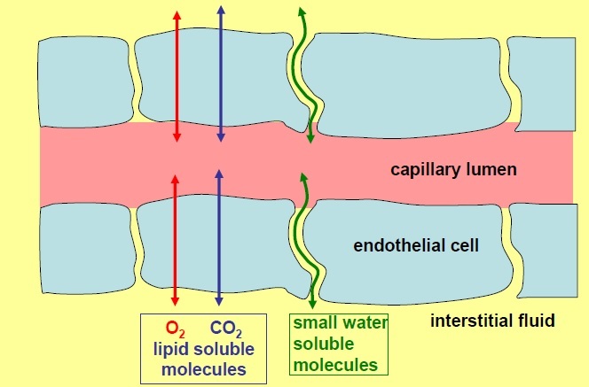
98% of exchange is by diffusion.

  • Capillaries are composed of a single layer of endothelial cells surrounded by basal lamina.
  • Lipophilic molecules such as O2 and CO2 can diffuse directly through the lipid blayer
  • Small, hydrophilic molecules such as glucose, amino acids and lactate diffuse through small pores between endothelial cells.
  • All molecules will move down their concentration gradient
3
Q

What does the rate of diffusion depend on?

A
  • Area
  • Diffusion Resistance
  • Concentration Gradient (difference)
4
Q

Explain how the rate of diffusion depends on the area available for exchange

A
  • The larger the area, the faster the rate of diffusion
  • Area for exchange between capillaries and tissues is generally very large - depends on CAPILLRY DENSITY (number of capillaries per unit volume)
  • A tissue which is more metabolically active will have more capillaries.
  • Not all capillaries are always perfused so it represents the maximum area for exchange
  • Generally, however, area is not limiting factor
5
Q

Explain how the rate of diffusion depends on the diffusion resistance

A

the difficulty of movement through the barrier. Depends on:

  • Nature of the molecule: hydrophilic or lipohilic, size (small hydropilic molecules can diffuse readily through pores, large hydrohilic molecules may be too big)
  • Nature of the barrier: e.g.pore size and number of pores for hydropilic substances (some tissues have more or larger pores). Tight junctions e.g. in the brain decreases pore size which limits diffusion.
  • Path length (depends on capillary density; path is shortest in the most active tisssues (highest capillary density)
  • Diffusion resistance is mostly low - not generally the limiting factor.
6
Q

Explain how the rate of diffusion depends on the concentration gradient

A
  • The greater the concentration gradient, the greater the rate of diffusion
  • The concentration which matters is between capillary blood and tissues
  • FOr exchange to continue, the concentration gradient between the capillary blood and tissues must be maintained.
  • Unless blood is supplied at an appropiate rate, the gradients driving exchange will dissipate and the nutrients will not be supplied at the right rate.
  • A substance which is used by the tissues will have a lower concentration in capillary blood than arterial blood e.g. O2. How much lower depends on RATE tissues use the substance (causes [O2] drop) and RATE of blood through the capillary bed (if blood flow is fast, [O2] is maintained)
7
Q

What does the suply of materials to the tissues depend most critically on?

A
  • Concentration gradients driving xchange
  • If all other things are equal, the supply of nutrient to a tissue depends most crtically on maintaining the right FLOW of bloodfor the prevailing level of metabolic activity. The CVS must maintain appropriate flows through ALL tissues.
8
Q

What is the rate of blood flow also known as?

A

The perfusion rate which can be constant or vary enormously.

9
Q

Describe the blood flow to the Brain

A

Metabolic needs are constant and can be met by a flow of 0.5ml/min/g

Th brain is extremely intolerant of flow interruption - blood flow is high and constant - 750ml/minute

10
Q

Describe the blood flow to the heart

A
  • At rest the heart needs 0.9ml/min/g but if the heart has to work hard this may increase four fold.
  • The heart is also extremely intolerant of inadequate flow.
  • Minimum: 300ml/min
  • Maximum: 1200ml/min
11
Q

Describe the blood flow to the kidney

A
  • Requires high constant flow to maintain its function (3.5ml/min/g) though most flow is not nutritive.
  • Minimum: 1200ml/min
  • Maximum: 1200ml/min
12
Q

Describe the blood flow to the Gut and Liver

A
  • They are connected in series via the hepatic portal system
  • At rest they receive 1ml/min/g
  • Digestion of meal generates a substantial increase in flow.
  • Short term flow reduction is tolerable.
  • Minimum: 1400ml/min
  • Maximum: 2400ml/min
13
Q

Describe the blood flow to the skin

A
  • Skin is not metabolically very active and may be upported by 0.01ml/mi/g, though flow may increase to 1.5-2ml/min/g for thermoregulation
  • Minimum: 200ml/min
  • Maximum: 2500ml/min
14
Q

Describe the blood flow to the rest of the body

A
  • A fairly constant demand: 200m/min
15
Q

What is the total body blood flow?

A
  • At rest: 5L/min
  • In exercise: 25L/min
  • CVS must deliver the above and maintain a blood flow of 750ml/min to the brain at all times.
  • It must also maintain a blood flow to heart and kidneys at al times.
  • Blood flow to skeletal muscle can be very high during exercise and gut blood flow is high after a meal
16
Q

How do you regulate Blood flow (so that cardiac output of the pump is distributed appropriately)?

A
  • add resistance to the system so that blood doesn’t only flow to the parts that are the easiest to perfuse (brain is harder to perfuse due to gravity)
  • Reduce the ease with which some regions are perfused in order to direct blood flow to the more difficult to perfuse regions
  • Arterioles and pre-capillary sphincters are the resistance vessels.
17
Q

Explan the direction of the blood

A
  • Heart pumps blood –> arteries supply –>. arterioles supply –> capillaries drain into –> venules drain into –> veins return blood –> heart
  • Heart is two pumps in series. The left heart pumps blood around the systemic circulation. The right heart pumps blood around the pulmonary circulation.
18
Q

The total flow in the system has to be able to change. What does this require?

A
  • A temporary store of blood which can be returned to the heart at a different rate.
  • Veins have thin walls which can easily distend or collapse enabling them to act as a variable reservoir for blood.
  • The reservoir can then be called up to cope with temporary imbalances between the amount of blood returning to the heart and the amount that it is required to pump out.
  • This store is in the veins.
19
Q

Explain the distributon of blood in the CVS

A
  • 67% veins
  • 11% arteries anD arterioles
  • 5% capillaries
  • 17% heart and lungs
20
Q

Where is blood flow fastest?

A
  • Where the total cross sectional area is least:
  • Cross sectnal area of aorta = 2.5cm(2) so fast flow (as the arteries branch, the total cross sectional area of the vascular bed increases)
  • Total cross setional area of capillries = 4500cm(2) so slow flow)
  • Combined cross-sectioal area of vena cavae = 8cm(2) so slightly faster flow
21
Q

Describe the great arteries arising from the heart

A
  • Carry blood flow away from the heart to the capillary beds
  • Pulmonary trunk arisng fom the right ventricle. It bifurcates into the right and left pulmonary arteries that enter the lungs
  • Aorta is the major artery arising from the left ventricle. It courses in a posteriorly oblique arch to descend into the thoracic cavity.
  • 3 main arterial trunks: the brachiocephalic artery, the left common carotid artery and the left subclavian artery arise from the arch of the aorta. They are elastic arteries
22
Q

What are the 3 Major Types of Arteries?

A
  • Elastic conducting arteries (widest diameter)
  • Muscular distributing arteries (intermediate diameter)
  • Arterioles (narrowest)
23
Q

Describe Elastic Arteries

A
  • Systole: LV contraction causes the blood pressure n the aorta to rise to ~120mHg. The walls of the elastic aorta and other elastic arteries stretch.
  • Diastol: aorti semilunar valve closes. Walls of aorta recoil maintaining pressure on the blood and moving it towards the smaller vessel. Aortic pressure drops to 70-80mmHg (diastolic)
  • So elastic arteries conduct blood away from the heart but also act as pressure reservoirs. During diastole they act as auxiliary pumps giving back the elastic energy stored during systole.
24
Q

Describe the Characteristics of Elastic Arteries

A
  • Tunica intima: endothelial cells with long axis orientated parallel to the long axis of artery. Narrow subendothelium of connective tissue with discontinuous internal elastic lamina
  • Tunica media: 40-70 fenestrated (contains holes) elasticmembranes. Smooth muscle cells (producing collagen , elastin and extracellular matrix) between these lamellae. Thin external elastic lamina may be present. Branching elastic lamellae.
  • Tunica adventitia: thin layer of fibroelastic connective tissue (contains collagen), vasa vasorum (‘vessels of vessels’), lymphatic vesels and nerve fibres (has got neurovascular supply)
25
Q

What are the three layers in the walls of arteries and veins?

A
  • Tunica intima next to the lumen: endothelium and subendothelial layer
  • Tunica media (intermediate layer)
  • Tunica adventitia (outer layer)
  • In arteries: internal elastic lamina between tunica intima and tunica media, external elastic lamina between tunica media and tunica adventitia.
26
Q

Describe the characeristics of Muscular Arteries (Distributing)

A
  • Tunica Intima: endothelium, subendothelial layer, thick internal elastic lamina
  • Tunica media: 40 circularly arranged layers of smooth muscle cells. These cells are connected by gap junctins for coordinated contraction. Prominent external elastic lamina
  • Tunica adventitia: thin layer of fibroelastic connective tissue containing vasa vasorum, lymphatic vessels and nerve fibres (unmyelinatd nerve endings)
27
Q

Describe Muscular arteries

A
  • Vasoconstriction is stimulated by sympathetic nerve fibres
  • Neurotransmitter Noradrenaline released at the nerve endings diffuses through fenestrations in te external elastic lamina into the external tunica media to depolarise some of the superficial smooth muscle cells.
  • As arteries diminish in diameter, the number of smooth muscle layers in the tunica media diminishes.
28
Q

What is an end artery?

A
  • Terminal artery supplying all or most of the blood to a body part without significant collateral circulation
  • Such arteries undergo progressive branching without the development of channels connecting with othe arteries, so that, if occluded, there is insufficient blood supply to the dependant tissue.
  • Examples include the coronary artery, splenic artery and renal artery.
  • Best examples of absolute end arteries (anatomically true end arteries) are the central artery to the retina and the labyrinthine artery of the internal ear.
29
Q

What is Bridging?

A

Compression of a segment of a coronary artery during systole, resulting in narrowing that reverse during diastole.

30
Q

What are arterioles?

A
  • Diameter of less than 0.1mm
  • 1-3 layers of smooth mucle in tunia media
  • Thin internal elastic lamina is present in larger arterioles only.
  • In small arterioles the tunica media is composed of a single smooth muscle cell that completely encircles the endothelial cells
  • The external elastic lamina is absent
  • The tunica adventitia is scant.
  • Function: to regulate the amount of blood reaching an organ or tissue and more generally in regulating blood pressure.
31
Q

What are metarterioles?

A
  • Arteries that supply blood to capillay beds are called metarterioles
  • They differ from arterioles in that the smooth muscle layer is not continuous.
  • Rather the individual muscle cells are spaced apart and each encircles the endothelium of a capillary arising from the metarteriole. This is a PRECAPILLARY SPHINCTER
  • Each smooth muscle cell is believed to function as a sphincter (upon contraction, controlling blood flow into the capillary bed)
32
Q

What do precapillary sphincters do?

A
  • They allow the arterioles and metarterioles to serve as flow regulators for the capillary beds.
  • When the precapillary sphincters are open, there is plentiful blood flow through the capillary bed.
  • When the precapillary sphincters are closed, the blood flow through the capillary bed is greatly reduced.
33
Q

Describe arteriole dilation and constriction

A
  • most arterioles can dilate to 60-100% of their resting diameter, or maintain up to a 40% constriction for a long time. This regulation directs blood where it may most be needed.
  • During strenuous physical exertion, blood flow to skeletal muscles is increased by dilation of arterioles and blood flow to the intestines is decreased by constriction of arterioles.
  • After ingeston of a large meal, the reverse is true
34
Q

What do lymphatic capillaries do?

A

drain away excess extracellular fluid, returning it to the blood at the junctions of the internal jugular and subclavian veins.

35
Q

What happens at the capillaries?

A
  • Passing RBCs fill virtually the entire capillary lumen, minimizing the diffusion path to adjacent tisses.
  • It is during passage through the capillaries that blood velocity is at its lowest (0.3mm/s), allowing time for gas and nutrient exchange with surrounding tissues.
  • 7-10 micrometres in diameter, usually less than 1m long
  • Essentially a tube just large enough to allow the passage of blood cells one a a time]
  • Made up of a single layer of endothelium and its basement membrane
36
Q

What are the three types of Capillaries?

A
  • Continuous
  • Fenestrated
  • Sinusoidal or discontinuous (Sinusoids)
37
Q

Describe Continuous Capillary

A
  • Most common type, located in nervous, muscle and onnective tissue, exocrine glands and the lungs
  • Continuous endthelial layer - cells joined by tight or occludng junctions
  • Pericytes form a branching network on the outer surface of the endothelium. These cells are capable of dividing into muscle cells or fibroblasts, during angiogenesis, tumour growth and wound healing.
38
Q

Describe Fenestrated Capillaries

A
  • Found in parts of gut, endocrine glands and renal glomerulus
  • little ‘windows’ or interruptions exist across thin part of the endothelium; these are bridged by thin diaphragm (except the renal glomerulus)
39
Q

What are 4 possible routes of transport across the endothelial wall of a fenestrated capillary?

A
  • Direct diffusion
  • Diffusion through intercellular cleft
  • Diffusion through fenestration (‘pore’)
  • Or via Pinocytotic vesicles
40
Q

Describe Sinusoidal/Discontinuous Capillaries

A
  • Larger diameter (30-40 micrometres) and slower blod flow
  • Seen in liver, spleen and bone marro
  • Gaps exist in the walls allowing whole cells to move btween blood and tissue (under certain conditions, some blood cells leave the circulatory system to enter the tissue spaces)
  • Sinusoids may contain special lining cells and an incomplete basal lamina + generally larger in diameter compared to the other capillaries.
41
Q

Describe Postcapillary Venules

A
  • They receive blood from capillaries
  • Diameter 10-30 micrometres
  • More permeable than capillaries
  • They have lower pressure than that of capillaries or the surrounding tissue so fluid tends to drain into them except when an inflammatory response is operating (in which case fluid and leukocytes emigrate)
  • These venules are the preferred location for emigration of leukocytes from the blood.
42
Q

Describe Venules

A
  • As the diameter of the merging venules increases to more than 50 micrometres, smooth muscle fibres begin to be associated with the endothelium - a tunica media appears (the endothelium is associated with pericytes or thin smooth muscle cells to form a very thin wall).
  • A valve in a venule consists of thin intimal extensions. By pressing these together, these restrict retrograde flow of blood.
43
Q

Describe Veins

A
  • as a general rule, veins have a larger diameter (larger lumen) than any accompanying artery, and a thinnet all that has more connective tissue and fewer elastic and muscle fibres.
  • Small and medium sized veins have a large well-developed adventitia layer. The tunica intima is thin, as is the tunica meda (2 or 3 layers of smooth muscle)
  • Large eis have diameters >10mm. The tunica intima is thicker. Most large veins do not have a prominent tunica media but have a well-developed tunica adventita.
  • A excepton is the superficial veins of the legs, which have a well-defined muscular wall, possibly to resist distension caused by gravity
44
Q

Describe valves in veins, and how is blood flow maintained?

A
  • Semilunar paired valves act, together with muscle contraction to propel blood towards the heart (one direction)
  • The veins collapse if blood pressure is not maintained; the blood flow in arteries is the result of cardiac systolic pressure whereas blood flow in veins is, to a great extent, detemined by the “muscle-pump” action in the leg and pressure factors in the abdominal and thoracic cavities.
45
Q

What are Venae Comitantes?

A
  • Vena comitans (singular) but normally occur in pairs (venae comitantes)
  • Venae comitantes are the deep, paired veins that, in certain anatomical positions, accompany one of the smaller arteries on each side of the artery - close association.
  • The three vessels are wrapped together in one sheath.
  • The pulsing of the artery promotes venous return within the adjacent, parallel, paired veins
  • Example: brachial, ulnar and tibial venae comitantes.
46
Q

Describe large veins

A
  • well-developed longitudinally orientated smooth muscle in the tunica adventitia in addition to the circularly arranged smooth muscle in the tunica media
  • Examples: vena cavae and the pulmonary, portal, renal, internal jugular, iliac and azygous
47
Q

What is the Vasa Vasorum?

A
  • network of small blood vessels that supply larger vessels with blood.
  • The largest blood vessels in the body such as the aorta, depend on this support network to maintain healthy function.
  • Both oxygenated and deoxygenated blood are carried to and from larger essels by these tiny bloo vessels.
  • In order to effectively receive oxygen from the bloodsteam, cells must be very close to a blood vessel or capillary. Because the large veins and arteries are so thick, their outer and middle cell layers cannot be adequately nourished without vasa vasorum.
48
Q

Descrie the position of the heart in situ

A
  • snugly enclosed within the inferior mediastinum, the medial cavity of the thorax
  • Apex is directed towards the left hub and rests on the diaphragm, approximately at the level of the fifth intercostal space.
  • Broad posterosuperior aspect or base, from which the great vesels of the body emerge, point towards the right shoulder and lies beneath the second rib.
49
Q

Talk about the Gross Anatomy of the Heart

A
50
Q

Describe the first layer of the pericardium

A

Pericardium is a fibroserous membrane that covers the heart and the beginning of its great vessels.

  • Tough fibrous external layer
  • Continuous superiorly with the tunica adventitia of the great vessels entering and leaving, and with the pretacheal layer of deep cervical fascia.
  • Continuous inferiorly with the central tendon of the diaphragm
  • Pericardium is influenced by movements of the heart and great vessels, the sternum and diaphragm
  • The heart is relatively well tethered in place.
  • Pericardium protects heart against sudden overfilling because it is so inextensible and closely related to the great vessels that pierce it superiorly
51
Q

What is the pericardial cavity?

A

Potential space between opposing layers of the parietal and visceral layers of serous pericardium. Normally contains a thin film of fluid that enables the heart to move and beat in a frictionless environment as the serous pericardial layers slide smoothly across each other.

52
Q

Describe the parietal and visceral layers of the pericardium

A
  • Visceral layer of serous pericardium forms the epicardium, the outermost of three layrrs of the HEART WALL.
  • It extends onto the beginning of the great vessels, becoming continuou with the parietal layer of serous pericardium where the aorta and pulmonary trunk trunk leave the heart, and where the superior vena cava and inferior vena cava and pulmonry veins enter the heart.
  • The parietal layer lines the interior of the loosely-fitting fibrous pericardium.
53
Q

What is the Transverse Pericardial Sinus?

A

transversely running passage posterior to the intrapericardial parts of the pulmonar trunk and ascending aorta, anterior to the superior vena cava and superior to the atria of the heart.

clinically sigificant to cardiac surgeons as after opening the pericardial sac anteriorly, a finger can be passed through the transverse pericardial sinus posterior to the ascending aorta and pulmonary trunk. Surgeons can stop or divert the circulation of blood in these arteries during surgery.

54
Q

What is the Oblique Pericardial Sinus?

A
  • wide pocket-like recess in the pericardial cavity posterior to the base of the heart, formed by the left atrium.
  • Sinus can be entered inferiorly and will admi several fingers however they cannot pass around any of these structures because the sinus is a blind sac (cul-de-sac)
55
Q

Describe the arterial supply to the pericardium

A
  • Mainly from a slender banch of the internal thoracic artery, the pericardiacophrenic artery
  • Smaller contributions of the blood come from the musculophrenic artery (a terminal branch of the internal thoracic artery), bronchial, oephageal and superior phrenic arteries (branches of the thoracic aorta), coronary arteries (visceral layer of serous pericardium only)
56
Q

Describe the nerve supply of the pericardium

A
  • Phrenic nerves (C3-C5), primary source of sensory fibres; pain sensations conveyed by these nerves are commonly referred to the skin (C3-C5 dermatomes)
  • Vagus nerve (function uncertain)
  • Sympathetic trunks, vasomotor
57
Q

What is pericarditis?

A
  • Inflammation of the pericardium usually causes chest pain
  • Serous pericardial layers are rough - friction of the roughened surfaces may sound like the rustle of silk when listening with a stethoscope over the left sterna order and upper ribs (pericrial friction rub)
58
Q

What are consequences of pericarditis?

A
  • A chronically inflamed and thickened pericardium may calcify, seriously hampering cardiac efficiency.
  • Some inflammatory diseases produce pericardial effusion (passage of fluid from pericardial capillaries into the pericardial cavity or an accumulation of pus)
  • If extensive pericardial effusion exists, the compromised volume of the sac does not allow full expansion of the heart, limiting the amount of blood the heart can receive which in turn reduces cardiac output.
  • Pericardiocenetesis: drainage of fluid from the pericardial cavity is sometimes necessary
59
Q

What are the coronary arteries?

A
  • first branches of the aorta that supply the myocardium and epicardium
  • RIGHT and LEFT coronary arteries arise from the corresponding aortic sinuses at the proximal part of the ascending aorta, just superior to the aortic valve.
60
Q

Describe the right coronary artery (RCA)

A

Travels in the right atrioventricular groove, passing posteriorly beween the RA and the RV.

Supplies blood to the RV via acute marginal branches

Also supplies blood to the inferior and posterior walls of the ventricles and posterior one third of the interventricular septum after the distal RCA gives rise to a large branch, the posterior descending artery (in most people)

Just before giving off the posterior descending artery, the RCA usually gives off the AV nodal artery

In 70% of people, RCA also supplies SA node.

61
Q

Describe the Left Main Coronary Artery

A
  • Passes between the LA and the pulmnary trunk to reach the atrioventricular groove
  • There it divides into the Left Anterior Descending (LAD) coronary artery and the circumflex artery.
62
Q

Describe LAD

A
  • Travels within the anterior interventricular groove toward the cardiac apex.
  • It gives off septal branches which supply the anterior two thirds of the interventricular septum and the apical portion of the anterior papillary muscle.
  • Also supplies the anterior surface of LV via diagonal branches.
63
Q

Describe the Circumflex Artery

A
  • continues within the left AV groove and passes around the left border of th heart to reac hthe posterior surface.
  • It gives off large obtuse marginal branches that supply the lateral and posterior wall of the left ventricle.
64
Q

Describe the Venous Drainage of the heart

A
  • Drained mainly by veins thatempty nto the coronary sinus and partly by small veins that empty into the right atrum
  • Coronary sinus, the main vein of the heart, is a wide venous channel that runs from left to right in the posterior part of the coronary sulcus.
  • The coronary sinus receives the great cardiac vein at its left end and the middle cardiac vein and small cardiac veins at its right end
  • The smallest cardiac veins are minute vessels that begin in the capillary beds of the myocardium and open directly into the chambers of the heart, chiefly the atria. Although called veins, they are valveless communications with the capillary beds of the myocardium.