Session 4 Flashcards Preview

ESA2 CVS > Session 4 > Flashcards

Flashcards in Session 4 Deck (59)
Loading flashcards...
0
Q

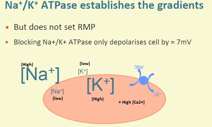
Does the Sodium Pump set the Resting Membrane Potential?

A

The Na+/K+-ATPase establishes the ionic gradients but does not set the resting membrane potential

Blocking the Na+/K+-ATPase only depolarises cell by approximately ~7mV.

1
Q

What sets the Resting Membrane Potential?

A

RMP is set largely due to K+ permeability of the cell membrane at rest. K+ channels (background inward rectifier type) are open at rest (high intracellular [K+] and low extracellular [K+]) so K+ ions move out of the cell down their concentration gradient.

The small movement of positively charged ions out of the cell makes the inside more negative with respect to the outside. As charge builds up, an electrical gradient is set up.

Resting membrane potential moves towards E(K)

2
Q

Why doesn’t RMP = E(K)

A

Very small permeability to other ion species at rest (other electrochemical gradients present)

3
Q

What effect do action potentials have on cardiac myocytes?

A

APs trigger increase in cytosolic [Ca2+]

A rise in calcium is required to allow actin and myosin interaction - generated tension

4
Q

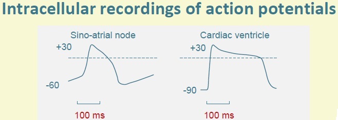
Draw the changes in membrane potential of cardiac ventricular and sinoatrial node pacemaker cells, including time durations

A
5
Q

Describe the membrane permeability changes and ionic currents underlying the ventricular (cardiac) action potential

A

Opening voltage-gated fast Na+ channels causes a large rapid upstroke leading to a Na+ influx which drives membrane potential to E(Na)

But E(Na) is not reached because of Na+ channel rapid inactivation and because of the permeability for other ions.

Initial depolarisation occurs - transient outward K+ current is caused by a specific type of K+ channels which open briefly before closing (voltage gated i(to)). The plateau region is caused by the opening of voltage-gated Ca2+ (L type) channels (some K+ channels also open).

The influx of [Ca2+] drives the membrane potential to E(Ca) (~+120mV) BUT this is not reached because of other open channels (K+). So the plateau region stays around 0mV - balanced with K+ efflux.

The downward stroke (repolarisation) is caused by the inactivation of Ca2+ channels (which take much longer to inactivate than Na+ channels) and the opening of voltage-gated K+ channels which allow an efflux of K+ and other K+ channels.

6
Q

What happens when the heart muscle is relaxed?

A

During diastole, the potential difference between inside and outside of the cells is negative inside. Except for the pacemaker cells, this potential difference is constant during diastole.

7
Q

Describe the cardiac action potential duration

A

About 280ms at rest because of the plateau sustained mainly by calcium channels.

Length of the AP is crucial; once the AP has begun in any part of the heart it needs to be long enough for the cell still to be depolarised when the last cell of the myocardium starts its zap. Therefore one AP in the pacemaker generates just one AP in every cell of the heart. This will produce a single heart beat.

8
Q

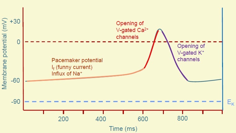
What is the Pacemaker Potential?

A

No stable resting membrane potential - just a long slow period of depolarisation (the diastolic period) which is called the pacemaker potential.

The pacemaker potential is the initial slow to threshold (I(f) funny current). It is activated by membrane potentials more negative than -50mV. The more negative the membrane potential, the more HCN channels are activated.

9
Q

What are HCN channels?

A

Hyperpolarization-activated Cyclic Nucleotide-gated Channels

Activated by Hyperpolarization

Activated by binding some molecules such as cAMP

Allows influx of Na+ channels Responsible for the pacemaker potential - the long period of slow depolarisation during diastole in SA node cells.

10
Q

Describe the SA Node action potential

A

After the pacemaker potential, the upstroke is caused by the opening of voltage-gated Ca2+ channels (fast Na+ channels are all in an inactive state due to the persistently less negative membrane voltage)

The downward stroke is (repolarisation) caused by the opening of voltage-gated K+ channels (K+ efflux) and closure of the Ca2+ channels.

The HCN channels open during the repolarisation of the cell as the potential approaches its most negative values.

11
Q

Describe Action Potentials throughout the heart

A

Action potential varies throughout the heartheart.

The sinoatrial node is fastest to depolarise so it sets the rhythm (is the pacemaker) but OTHER PARTS OF THE CONDUCTING SYSTEM (e.g.purkinje fibres, AV node) also have automaticity but they are slower.

12
Q

Describe the characteristics of cardiac myocytes

A

Single central nucleus

Cells joined together at intercalated discs

Gap junctions found on intercalated discs (large ion channels formed by connexon proteins) are NON-SELECTIVE - permeable to a wide range of ions. Gap junctions permit movement of ions and electrically couple cells (movement of positively charged ions pout of cell causes depolarisation)

Desmosomes are also found on intercalated disks - RIVET cells together. Cells are mechanically tethered together to contract together.

13
Q

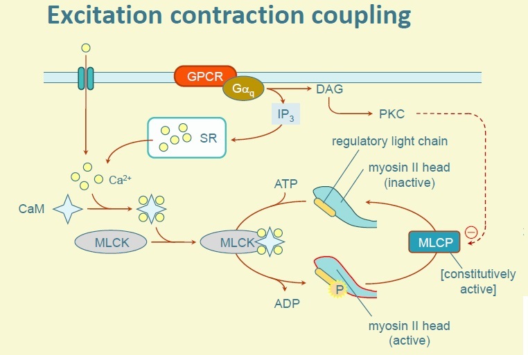
Describe how the intracellular [Ca2+] increases

A

Depolarisation opens L-type Ca2+ channels in T Tubule system

Localised Ca2+ entry (via the L-type channels) opens Calcium-Induced Calcium Release (CICR) channels in the sarcoplasmic reticulum.

Close link between the L-type channels and Ca2+ release channels. 25% of intracellular [Ca2+] enters across sarcolemma (L-type channels) and 75% of intracellular [Ca2+] is released from SR, causing the intracellular [Ca2+] to rise during the plateau phase.

Release of Ca2+ can’t take place until Ca2+ enters across sarcolemma first. Ca2+ binds to TnC subunit causing a conformational shift so Tropomyosin moves away and unblocked the myosin binding site on the actin filament.

14
Q

What is the Sliding Filament Theory?

A

1) Attachment: myosin head tightly bound to actin molecule.
2) Release: ATP binds to the myosin head causing it to uncouple from the actin filament.
3) Bending: hydrolysis of the ATP caused the uncoupled myosin head to bend and advance a short distance (5nm)
4) Force Generation: the myosin head binds weakly to the actin filament causing release of inorganic phosphate which strengthens binding and causes the power stroke in which the myosin head returns to its former position.
5) Reattachment: ATP binds to the myosin head causing detachment from actin. The cycle will repeat.

Individual myosin heads attach and flex at different times causing movement. Muscle shortens as the thick and thin filaments slide past each other.

15
Q

Describe Relaxation in Cardiac Myocytes

A

Intracellular [Ca2+] must return to resting levels in order for relaxation to take place.

Most is pumped back into the SR by SERCA which is stimulated by raised Ca2+ levels.

Some Ca2+ exits across cell membrane via Sarcolemmal Ca2+ ATPase and Na+/Ca2+ Exchanger,

In the heart, the force generated in a cell, at a given degree of stretch is proportional to Ca2+ concentration. Therefore the force of contraction depends upon the balance between the rate of entry of Ca2+ into the cytoplasm and its rate of removal. As the plateau phase is long, the muscular contraction is sustained for 200-300ms which is essential for the normal pumping activity of the heart.

16
Q

How is tone of blood vessels controlled?

A

Vascular smooth muscle within the tunica media is not striated.

Contraction still takes place but myofilaments are arranged differently.

Gap junctions are still present (aqueous pores allowing ion movement) and they electrically couple cells.

Contraction of these vascular smooth muscles leads to an increased tone of the vessel, narrowing the lumen.

17
Q

How does binding of Ca2+ to Calmodulin control excitation coupling?

A

Binding of Ca2+ to Calmodulin activates Myosin Light Chain Kinase (MLCK) which phosphorylates the myosin light chain to permit interaction with actin.

Myosin Light Chain must be activated (via phosphorylation by MLCK) to enable actin-myosin interaction which in turn leads to contraction.

Relaxation occurs as Ca2+ levels decline.

Phosphorylating by Protein Kinase A inhibits MLCK therefore inhibiting contraction.

18
Q

Why is the Pacemaker Action Potential different to the Cardiac Action Potential?

A

Pacemakers initiate the AP themselves rather than by conduction of excitation from surrounding cells. In diastole, the membrane potential is unstable - depolarises steadily (pacemaker potential).

This is thought to be due to the HCN channels that are permeable to Na+ ions which are different to the fast Na+ channels - they are activated by Hyperpolarization, referred to as the funny current. Pacemaker cells do not have fast Na+ channels but as the membrane depolarises with the pacemaker potential, voltage gated Ca2+ channels eventually open producing a faster rate of depolarisation to a positive membrane potential.

The opening of these Ca2+ channels is not sustained - there is no plateau and the action potential is triangular in shape.

As soon as the membrane is repolarised back to -600mV, it begins to depolarise again slowly - the next pacemaker potential begins until threshold is reached again and the next AP occurs.

19
Q

Describe the Action Potential in the Conducting Fibres

A

Purkinje fibres conduct excitation through the ventricular myocardium.

They have long APs but within the AV node and the bundle of His, there are cells capable of pacemaker activity.

Their natural rate is much slower than the SA node so they are normally overridden.

If however there is a conduction block, they may become important.

20
Q

The ANS is an efferent system. It is important for regulation many physiological functions - what physiological functions?

A

Heart rate,

blood pressure,

body temperature etc (homeostasis - keeping everything in balance)

Coordinating the body’s response to exercise and stress

Largely outside voluntary control

21
Q

Give examples of what the ANS exerts control over

A

Smooth muscle (vascular and visceral)

Exocrine secretion e.g. Saliva and sweat

Rate and force of contraction in the heart

22
Q

Describe the divisions of the ANS

A

Based on anatomical grounds: Parasympathetic nervous system Sympathetic nervous system

NB: some textbooks include a third division: the enteric nervous system (network of neurones surrounding GI tract is normally controlled via sympathetic and parasympathetic fibres)

23
Q

Describe the organisation of sympathetic and parasympathetic systems (generally)

A

Two neurones arranged in series

Cell body of Preganglionic neurone in the CNS

Other neurone is located entirely outside of the CNS

Cell bodies of these neurones are located in structures knwon as ganglia

Preganglionic fibres leave the CNS and then synapse with post-ganglionic cell bodies in the ganglia.

Post-ganglionic fibres run to the innervated structures and form neuro effect junctions with effector cells

Transmission at both ganglionic synapses and neuro effector junctions is by the release of chemical messengers - neurotransmitters.

24
Q

Describe the Organisation of the Sympathetic Division

A

Thoraco lumbar origin: Preganglionic neurones arise from segments T1 to L2 (or L3)

Preganglionic fibres are SHORT

Postganglionic fibres are LONG

Most synapses occur in the paravertebral chain of ganglia. The chain extends further cranially and caudally than the origins of the nerves so neurones can travel up or down before synapses. Some neurones don’t synapse in the chain but synapse in a number of prevertebral ganglia: celiac, superior mesenteric, inferior mesenteric ganglia

Some ganglia are located in the neck and abdomen and these have longer pre-ganglionic fibres.

25
Q

Describe the Organisation of the Parasympathetic Division

A

Craniosacral origin (directly from the brain or sacral region)

Preganglionic fibres travel in cranial nerves (III, VII, XI and X) or Sacral Outflow from S2-S4

Long pre ganglionic fibres

Short post ganglionic fibres - synapses occur close to the target tissues or sometimes within the structures controlled by the system, There are however some post-ganglia in the neck and abdomen, located further away from the target organs.

26
Q

What do Preganglionic neurones of both divisions release?

A

Acetylcholine ACh binds to nicotinic ACh receptors on the post ganglionic cell.

Nicotinic ACh receptors have an ion channel, that when activated, allow an influx of Na+ and an efflux of K+ which leads to depolarisation and AP generation and propagation down the neurone.

Nicotinic ACh receptors are ligand gated.

27
Q

What are Postganglionic Sympathetic Neurones like?

A

Usually Noradrenergic - release noradrenaline.

The exception is sympathetic innervation of the sweat glands; here Postganglionic neurones release ACh which acts on Muscarinic ACh receptors.

28
Q

What are Postganglionic Parasympathetic Neurones like?

A

Most but not all are cholinergic - release ACh as neurotransmitter which act on Muscarinic ACh receptors on the effector cell

29
Q

Explain the Sympathetic nervous system innervation to the adrenal medulla

A

Preganglionic fibres run to the adrenal medulla which is made of modified (specialised) post-ganglionic cells, Chromaffin cells, which release adrenaline into the blood where it circulates throughout the body.

Circulating adrenaline will also act upon receptors in tissues, producing a more generalised effect.

30
Q

Give some examples of ANS Control: to the pupil of the eye

A

Sympathetic effect: dilation (contracts radial muscle); receptor: alpha1

Parasympathetic effect: contraction (contracts sphincter muscle); receptor: muscarinic M3

31
Q

Give some examples of ANS Control: airways of the lungs

A

Sympathetic effect: Relax; Receptor: Beta2

Parasympathetic effect: Contract; Receptor: M3

32
Q

Give some examples of ANS Control: to the heart

A

Sympathetic effect: increase rate and force of contraction; receptor: Beta1

Parasympathetic effect: decrease rate; receptor: M2

33
Q

Give some examples of ANS Control: sweat glands

A

Sympathetic Effect: Localised Secretion (e.g. Palms) receptor: alpha1; Generalised Secretion receptor: M3

NO PARASYMPATHETIC EFFECT

34
Q

Describe the receptors Noradrenaline and Adrenaline act on

A

Adrenoceptors: G-Protein coupled receptors with no integral ion channel

Types and Subtypes: alpha1, alpha2, beta1, beta2

Different tissues can have different subtypes with different signalling mechanisms, allowing for diversity of action and selectivity of drug action

Broadly speaking, alpha receptors are found on vascular smooth muscle. beta-receptors are found in the heart, smooth muscle of the airways of the lungs, adipose tissue and some blood vessels particularly in skeletal muscle.

35
Q

What are muscarinic receptors?

A

G-Protein Coupled Receptors (M1, M2, M3) with no integral ion channel

Parasympathetic post ganglionic neurones use ACh as a neurotransmitter.

ACh acts at muscarinic receptors on the effector cells.

36
Q

What does the Autonomic Nervous System do?

A

Regulates physiological functions

Where parasympathetic and sympathetic divisions both innervate a tissue they often have opposite effects

Sympathetic activity is increased under stress

Parasympathetic system is more dominant under basal conditions

Both work together to maintain balance

37
Q

How is sympathetic drive to different tissues regulated?

A

Independently so sympathetic activity to the heart can be increased without increasing activity to GI tract

On some occasions (fight or flight) there can be a more co-ordinated sympathetic response

38
Q

What does the ANS control in the Cardiovascular system?

A

Heart rate

Force of contraction of heart

Peripheral resistance of blood vessels

39
Q

Describe Parasympathetic input to the heart

A

Preganglionic fibres are derived from the 10th (X) Cranial Nerve (Vagal Nerve)

Synapse with post ganglionic cells on epicardium surface or within walls of heart (concentrated at SA and AV node)

Postganglionic cells release ACh which act on M2 receptors to DECREASE heart rate (-ve chronotropy) and decrease AV node conduction velocity

40
Q

Describe Sympathetic input to the heart

A

Postganglionic fibres from the sympathetic trunk innervates SA node, AV node and myocardium - releases Noradrenaline.

NA acts on B1 adrenoceptors: increases heart rate (+ve chronotropy) and increases force of contraction (+ve inotropy)

41
Q

What are the effects of Sympathetic activity on Pacemaker potentials?

A

Increases slope - makes pacemaker potential more steep and speeds up depolarisation so threshold is reached sooner and reduces time between action potentials.

Increases cAMP –> increases Heart Rate

42
Q

What are effects of Parasympathetic activity on the Pacemaker potential?

A

Increases K+ conductance (leading to Hyperpolarization) and decreases production of cAMP (leading to less activation of HCN channels)

Takes longer to reach threshold (Hyperpolarization) –> slows down Heart rate

NOTE: decrease in cAMP is more important than K+ conductance resulting in heart rate slowing down

43
Q

How does noradrenaline increase the force of contraction?

A

NA acting on B1 receptors in myocardium causes an increase in cAMP production

cAMP phosphorylates (activates) Ca2+ channels leading to increased Ca2+ entry during AP.

Increased uptake of Ca2+ in SR leads to increase in intracellular Ca2+ store - increased amount of available Ca2+

This leads to increased sensitivity of contractile machinery to Ca2+.

All this results in increased force of contraction - no opposite effect by the parasympathetic nervous system.

44
Q

Describe how the SNS innervation and effects on vasculature

A

The smooth muscle in the walls of arteries, arterioles and veins is innervated by the SNS.

Except in specialised vessels, sympathetic activity causes constriction of arterioles (vasoconstriction mediated by alpha1-adrenoceptors).

There is constant activity in the SNS (sympathetic vasomotor tone) tending to make arteriolar smooth muscle contract.

Tone varies from organ to organ e.g. In skin, vasomotor tone is high so arterioles, pre-capillary sphincters and arterio-venous anastomoses are generally shut down.

45
Q

Describe sympathetic outflow to various organs

A

Variation in sympathetic outflow produces large changed in skin blood flow, usually for the purposes of thermoregulation.

In skeletal muscles, vasomotor tone is high at rest but in exercise is antagonised by local release of vasodilator metabolites but also to a smaller extent by specialised vasodilator response to circulating levels of adrenaline (mediated through beta2-adrenoceptors)

In the gut vasomotor activity is high until after a meal is consumed when it is antagonised by various vasodilator substances produced in gut tissue.

Circulation in the brain is virtually unaffected by sympathetic activity.

46
Q

What type of adrenoceptors does most vasculature have?

A

Alpha1

But some blood vessels have beta2 adrenoceptors as well; skeletal muscle, myocardium and liver.

Circulating adrenaline has a higher affinity for beta2 adrenoceptors.

47
Q

What is the interplay between sympathetic vasoconstrictor tone and the action of vasodilator substances important?

A

It is the principle means by which distribution of blood flow around the CVS is controlled.

Vascular tone in skin and skeletal muscle is also a mechanism for controlling total peripheral resistance.

Sympathetic outflow to blood vessels is controlled from the brainstem - via the vasomotor centres in the medulla oblongata.

NOTE: sympathetic activity also produces veno-constriction which increases venous pressure, promoting return of blood back towards the heart.

48
Q

How does the PNS act on specialised blood vessels?

A

The PNS can act on specialised blood vessels such as in erectile tissue to cause vasodilation however it indirectly causes vasodilation in organs such as the gut by its stimulating activity of the organ which in turn causes the release of local vasodilator mediators.

49
Q

How does the PNS acting on beta2-adrenoceptors cause vasodilation?

A

Increases cAMP –> opens a type of potassium channel –> relaxation of smooth muscle.

50
Q

How does the SNS activating alpha1 adrenoceptors cause vasoconstriction?

A

Increase in intracellular [Ca2+] from stores and influx of extracellular Ca2+ –> contraction of smooth muscle.

Depolarisation also occurs.

51
Q

What is the role of local metabolites?

A

Active tissue produces more metabolites e.g. Adenosine, K+, H+, increased pCO2

Local increases in metabolites have a vasodilator effect.

More important for ensuring adequate perfusion of skeletal and coronary muscle than activation of Beta2-receptors.

52
Q

Describe the overall control of CVS

A

Changes in the state of the system are communicated to the brain via afferent nerves

Baroreceptors (high pressure side of system)

Atrial receptors (low pressure side of system)

The CVS control centres in the brain alters activity of efferent muscles - influences sympathetic output to the heart and blood vessels and Vagal outflow to nerve.

53
Q

Describe the effects of sympathetic and parasympathetic innervation to the heart

A

Both SA and AV nodes are invested by both systems. If all autonomic inputs are pharmacologically blocked, the intrinsic HR is about 100bpm.

The normal resting HR of 60bpm is produced because the parasympathetic system dominates at rest.

Both parasympathetic and sympathetic outflow to the heart are controlled by centres in the medulla oblongata which receives information from sensory receptors detecting blood pressure ‘baroreceptors’ and higher centres in the CNS.

Sympathetic nerve fibres also innervate ventricular cardiac myocytes - sympathetic activity increases the force of contraction of the heart muscle for a given fibre length.

Adrenaline from the adrenal medulla also acts on the heart.

The ANS provides the CVS control centre in the brainstem with means to control total peripheral resistance, distribution of blood flow and cardiac output.

54
Q

How is the action of the PNS on HR mediated? How is the action of SNS on HR and force of contraction mediated?

A

Action of the PNS on HR is mediated via ACh acting on M2 receptors.

Action of the SNS on the HR and force of contraction is mediated via NA acting in B1 adrenoceptors.

55
Q

What are Baroreceptors?

A

Nerve endings in the carotid sinus and aortic arch which are sensitive to stretch: increased arterial pressure stretches these receptors.

Baroreceptors sends afferent fibres along glossopharyngeal nerves to the CVS control centre in the medulla.

Bradycardia and vasodilation counteract increased mean arterial pressure.

56
Q

What are Sympathomimetrics?

A

Alpha-adrenoceptor agonists and Beta-adrenoceptor agonists (mimic action of sympathetic nervous system)

Cardiovascular uses: Administration of adrenaline to restore function in cardiac arrest Beta1 agonist: dobutamine may be given in cardiogenic shock (pump failure)

Adrenaline administered for anaphylactic shock (massive vasodilation occurs so can cause a low BP so adrenaline raised BP)

57
Q

What are Cholinergics?

A

Muscarinic agonists e.g. Pilocarpine is used in treatment of glaucoma (activates constrictor pupillae muscle)

Muscarinic antagonists e.g. Atropine or Tropicamide - increases heart rate, bronchial dilation - used to dilate pupils for examination of the eye.

58
Q

What are adrenoceptor antagonists?

A

Blocks effects of noradrenaline and adrenaline

Alpha1 adrenoreceptor antagonists: e.g. Prazosin is an anti-hypertensive agent - inhibits NA action on vascular smooth muscle alpha1 receptors - vasodilation

Beta-adrenoreceptor antagonists: E.g. Propranolol: non-selective B1/B2 antagonist. Slows heart rate and reduced forces of contraction (B1) but also acts on bronchial smooth muscle (B2) - bronchoconstriction E.g. Atenolol: selective B1 - less risk of bronchoconstriction (targets heart but not lungs)