Clinical Electrophysiology I Flashcards
(14 cards)
1
Q
Cellular Basis of Impulse Formation & Propagation
- Cardiac electrical activity
- Transmembrane potential
- Action potential
A
- Cardiac electrical activity
- Due to rapid changes in the electrical “charge” of the cell
- Caused by the rapid movement of + & - charged ions into & out of the cell
- Transmembrane potential
- “Ionic charge” of the cell
- Difference in potential voltage b/n the inside & outside of the cell
- Action potential
- Recording of the change in transmembrane potential over time
- Two patterns
- Classic: in myocytes
- Specialized: SA & AV nodes
- “All or none” response: signal begins when transmembrane potneital increases to threshold potential
- Changes in potential induce changes in adjacent cells to propagate the signal through the heart
2
Q
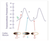
Classic APs: 5 Phases
A
- Phase 0: rapid depolarization
- Transmembrane potential crosses the threshold potential (-80 to -90 mV)
- Increased permeability to Na+ (& Ca2+)
- Increased influx of Na+ (& Ca2+) depolarizes the cell
- Makes the transmembrane potential positive
- Phase 1: rapid short repolarization
- Small efflux of K+
- Slightly decreases the positive charge of the cell
- Phase 2: plateau
- Balance b/n small inward (K+) & outward (Ca2+) currents
- Phase 3: repolarization
- Efflux of K+ makes transmembrane potential less +
- Phase 4: diastolic portion
- Decreased outward K+ current slowly increases + charge

3
Q
How Specialized APs Differ from Classic APs
A
- Phase 0: rapid depolarization
- Threshold potential = -40 mV
- Not as rapid b/c depolarization is due to Ca2+ influx, not Na+
- Phase 1: rapid short repolarization
- Absent
- Phase 2: plateau phase
- Absent
- Phase 3: repolarization
- Less rapid due to inactivation of Ca2+ channels & efflux of K+
- Phase 4: diastolic portion
- Steeper slope due to inward movement of Na+ ions (pacemaker current (If))
4
Q
Mechanisms of Rhythm Formation: Automaticity
- Automatic rhythms
- Most common example of automatic rhythm
- Phase 4
- Increasers of sinus rate
- Abnormalities in automaticity
A
- Automatic rhythms
- Characterized by gradual changes in rate
- Ex. Tachycardias: automatic warm-up & cool-down behavior
- Most common example of automatic rhythm: sinus rhythm
- SA & AV nodes are the only cardiac sites w/ intrinsic automaticity (pacemaker activity)
- Phase 4
- Enhanced diastolic potential currents (If)
- More positive slope
- Transmembrane potential increases to threshold potential sooner
- More APs in a given period of time –> faster rate
- Increasers of sinus rate
- Exercise
- Stress
- High catecholamine levels
- Excess thyroid hormone
- Abnormalities in automaticity
- Enhanced automaticity
- Increased automatic activity in cells w/ intrinsic activity (SA & AV nodes)
- Ex. inappropriate sinus tachycardia, junctional tachycardia
- Decreased automaticity
- More common
- Ex. tachy-brady syndrome, syndrome of chronotropic incompetence
- Automaticity in cells that don’t have intrinsic activity
- Ex. ectopic atrial tachycardia, accelerated idioventricular rhythms (slow ventricular tachycardia)
- Enhanced automaticity

5
Q
Mechanisms of Rhythm Formation: Triggered Activity (Triggered Automaticity)
- Triggered activity
- After potentials
A
- Triggered activity
- Pathological cellular depolarization that occurs spontaneously before another AP is expected
- After potentials (depolarizations) trigger depolarization of adjacent cells & propagate the cardiac impulse
- After potentials
- Early After Depolarizations (EADs)
- Occur soon after AP initiation (phase 2 or 3)
- Causes: acidosis, hypoxia, hypokalemia (metabolic abnormalities or ischemia)
- Delayed After Depolarizations (DADs)
- Occur later after AP initiatoin (phase 4)
- Causes: digitalis toxicity
- Responsible for physiological exercise-induced ventricular tachycardia
- Early After Depolarizations (EADs)

6
Q
Mechanisms of Rhythm Formation: Reentry
- Reentrant rhythms
- Responsible for…
- Only occur in the precence of 3 elements
A
- Reentrant rhythms
- Sudden initiation & termination of tachycardia
- Responsible for…
- The majority of supraventricular tachycardia (SVT) other than atrial fibrillation
- Nearly all ventricular tachycardia associated w/ coronary artery disease
- Only occur in the presence of 3 elements
- Conducting circuit: common upper & lower pathways, 2 separate limbs
- Different signal conduciton velocities int he two limbs: 1 fast, 1 slow
- Longer refractory period in the faster limb

7
Q
How Reentrant Elements Induce Rhythm Abnormality
- Normal situation
- Very early premature beat
- If a premature impulse is delivered a little later
A
- Normal situation
- Impulse –> upper common pathway –> 2 limbs
- Fast limb: moves so quickly that the impulse has time to be conducted up the slow pathway in a retrograde manner
- Slow limb: moves so slowly that there’s no evidence of limb conduction
- Normal impulse –> lower common pathway
- Impulse –> upper common pathway –> 2 limbs
- Very early premature beat
- Impulse –> upper common pathway –> partway through 2 limbs
- Limbs are both still within their refractory periods so can’t recover the ability to depolarize again
- If a premature impulse is delivered a little later
- Premature impulse –> upper common pathway –> 2 limbs
- Fast limb has a longer refractory period, so premature impulse is blocked
- Impulse is conducted in the slower limb
- Impulse –> lower common pathway –> conducted up fast limb in retrograde manner
- By the time the impulse –> upper common pathway, the fast limb has recovered from the refractory period
- Self-perpetuating cycle repeats –> tachycardia
- Premature impulse –> upper common pathway –> 2 limbs

8
Q
Cardiac Electrophysiology Studies of Reentrant Circuits
- Cardiac EP studies
- Reentrant circuit sizes
- Microscopic
- Intermediate size
- Macroscopic
A
- Cardiac EP studies
- Can start reentrant rhythms by adding timed premature impulses
- Reentrant circuit sizes
-
Microscopic: in reentrant atrial tachycardia
- Ex. sinus node reentry
-
Intermediate size: dual AV nodal physiology
- Responsible for AV node reentry tachycardia
-
Macroscopic: involves a large circuit of the atrium, AV node, ventricle, & accessory pathway
- Ex. AV reciprocating tachycardia associated w/ Wolff-Parkinson-White (WPW) Syndrome
-
Microscopic: in reentrant atrial tachycardia
9
Q
Anatomic Concepts in Cardiac Arrhythmias: SA Node
- Location
- Intrinsic pacemaker activity
- Innervation
- Impulse
A
- Location
- Heartbeat begins in the anterolateral junciton fo the RA & SVC
-
Intrinsic pacemaker activity: exhibits specialized APs that utilize the pacemaker (If) current
- Exhibits the highest/fastest activity that sets normal HR
- Innervation: right sided sympathetic & parasympathetic (vagus) nerves
- Parasympathetic tone predominates, so atropine (Ach agent) –> increased HR
- SA node impulse –> atrial tissue –> AV node
- Atrial tissue cells exhibit classic APs & don’t have intrinsic pacemaker activity
- Some specialized fibers may pass through the atrial tissue & provide preferential conduction to the AV node
- May play a role in some supraventricular tachycardia

10
Q
Anatomic Concepts in Cardiac Arrhythmias: AV Node
- Location
- Intrinsic pacemaker activity
- Innervation
- Delay
- Decremental conduction
- Impulse
- Electrical insulation
A
- Location
- Apex of triangle of Koch: septal attachment of the tricuspid valve + OS of the coronary sinus + tendon of Todaro
- Intrinsic pacemaker activity
- Slower rate than SA node
- Pace setting activity is suppressed by higher sinus rates
- Innervation
- Left sided sympathetic & parasympahtetic (vagus) nerves
- Delay
- Delays conduction of the signal from atrium to ventricle
- Responsible for majority of the delay in the PR interval
- When AV node is bypassed in teh presence of an accessory pathway (WPW syndrome), PR interval is shorter than normal
- Decremental conduction
- Slows conduction velocity w/ increasing impulse rates
- Governs ventricular rate: faster atiral rate –> slower signal conduction
- Helps control rapidity of ventricular rate in presence of rapid atrial rates
- Impulse
- Transmits impulse to Bundle of His
- Electrical insulation
- Cartilaginous structure that supports AV valves b/n atria & ventricles
- WPW syndrome: muscle fibers bridge this structure & provides a connection other than the AV node / His Bundle pathway

11
Q
Anatomic Concepts in Cardiac Arrhythmias: Bundle of His
- Location
- AV block
- Above the Bundle of His
- Below the Bundle of His
- Impulse
A
- Location
- Anatomical & electrical dividng point b/n ventricles & supraventricular structures
- Unifasicular
- Bifurcates into left & right bundle branches
- AV block
- Above the Bundle of His (in the AV node)
- Less worrisome, les slikely to be associated w/ fatal bradycardia
- Allows a higher rate escape rhythm
- Commonly Mobitz Type I block
- Congenital complete heart block: has an adequate ventricular response b/c the location is abov ethe Bundle of His
- Below the Bundle of His
- More worrisome
- Cells that can initiate ventricular contractoin don’t have intrinsic pacemaker activity to provide an adequate escape rhythm
- –> slow ventricular rate
- Above the Bundle of His (in the AV node)
- Impulse –> ventricualr myocytes w/ faster velocity than surrounding myocytes
- –> Right bundle
- Terminal named branch on the right side of hte heart
- –> Left bundle
- –> Anterior fascicle
- –> Posterior fascicle
- –> Right bundle
- Impulse –> purkinje fibers in ventricular tissue

12
Q
Consequences of Blocks in the Specialized Conduction System
A
- Rapid conduction through specialized conducting system allows all the ventricular myocytes to depolarize in a short period of time –> narrow QRS
- A block in this system causes the myocytes to rely on the passage of signals from cell to cell –> slower conduction velocity –> wider QRS
13
Q
Abnormal Anatomic Structures: Scar Tissue
- Surgical scars
- Surgical replacement & repair of heart valves
- Scars from a myocardial infarction
A
- Surgical scars
- Created during correction of congential heart disease
- Increase risk of atrial & ventricular arrhythmias
- Concern in surgically corrected Tetralogy of Fallot & congenital repairs that involve incisions or suturing into the atrial or ventricular tissue
- Surgical replacement & repair of heart valves
- Soruce of rhythm abnormalities
- Frequently takes form of heart block
- His Bundle: located near the noncoronray cusp of the aortic valve
- Surgery to this valve may cause postoperative conduction problems
- Mitral valve surgery may lead to conduction problems
- Esp if patient has calcification of the mitral valve annulus
- Scars from a myocardial infarction
- Scarred ventricle w/ multiple reentrant circuits & diverse conduction properties –> ventricular arrhythmias –> death
- Size of infarciton & extent of LV scarring is proportional to risk of sudden arrhythmic cardiac death
- Leads to indications for implantation of defibrillators

14
Q
Abnormal Anatomic Structures: Non-Scar Tissue
- Extra electrical connections b/n anatomic structures
- Extra connectoins b/n structures other than the atrium & ventricle
A
- Extra electrical connections b/n anatomic structures
- Wolff-Parkinson-White (WPW) syndrome: extra connection b/n atrium & ventricle (accessory pathway)
- Band of muscle fibers that bridges the AV node
- Allows the AV node to be bypassed
- Allows parts of the ventricular tissue to be depolarized before the signal passes through the AV / His Bundle system
- WPW syndrome –> supraventricular arrhythmias –> sudden arrhythmic death (w/ atrial fibrillation)
- Wolff-Parkinson-White (WPW) syndrome: extra connection b/n atrium & ventricle (accessory pathway)
- Extra connectoins b/n structures other than the atrium & ventricle
- Pre-excitation variants (nodofascicular / nodoventricular pathways) –> rhythm abnormalities
- Less common than WPW syndrome

