Local Control of Blood Pressure Flashcards
(13 cards)
1
Q
Blood flow to different regions
- During rest
- During non-rest
A
- During rest
- Total cardiac output = 6 L/min
- All tissues receive substantial perfusion
- Some vasoconstriction occurs because baseline SNS activity binds to alpha receptors on vascular smooth muscle
- Administration of a ganglionic blocker (ex. hexamethonium, a nicotonic receptor antagonist) decreases BP
- Some vasodilation occurs b/c smooth muscle (heart, skeletal muscles, liver) bind beta2 receptors
- Increases blood flow to heart, skeletal muscles, & liver
- beta2 receptors have a higher affinity for Epi than NE
- Vasodilation occurs in response to Epi release
- Some vasodilation occurs in the penis & clitoris during sexual excitement b/c sacral PNS neurons release nitric oxide
- During non-rest
- Total cardiac output increases
- Blood flow increases to some parts of the body & diminishes to others
-
Myogenic autoregulation: controls peripheral resistance by allowing vascular smooth muscle to regulate its own activity
- Influenced by release of paracrines: chemicals secreted by cells that affect contraction of nearby vascular muscle
- Also influenced by hormones
2
Q
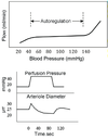
Myogenic Autoregulation
A
- Blood vessels adjust their diameter in response to alterations in BP so flow through the vascular bed remains constant
- Flow & perfusion pressure are directly proportional
- Vascular resistance increases proportionally to an increase in pressure
- Occurs in denervated vessels
- Neural influences aren’t needed to produce this effect
- All vascular beds autoregulate
- Increased pressure –> vascular smooth muscle cell walls stretch –> open stretch-sensitive Na+ channels –> depolarization –> open voltage-gated Ca2+ channels –> increase [Ca2+]i –> vasoconstriction
- Increased pressure –> initial distension of arteriolar wall is a stimulus for activation of smooth muscle cells –> vessel diameter decreases
- Autoregulation provides a constant rate of O2 delivery regardless of perfusion pressure

3
Q
Local control of vascular resistance
- Accumulation of chemicals during exercise
- Adenosine & K+
- Additional paracrine factors
A
- Accumulation of chemcials during exercise –> increased skeletal muscle perfusion
- Release of metabolites (H+ from acids, K+, CO2) –> vasodilation
- Low O2 levels –> vasodilation
- Mediated by release of adenosine from muscle cells during hypoxia
-
Adenosine & K+: metabolites w/ the strongest effects on vasoconstriction
- Increase adenosine –> increase cAMP –> activate protein kinase A (PKA) –> phosphorylate & open K-ATP channels –> K+ efflux –> hyperpolarization
- Increase [K+]o –> increase conductance through K-IR channel –> hyperpolarization
- Increase metabolism –> increase [K+]o –> open & increase K+ conductance throug hK-IR channels –> hyperpolarization –> close voltage-gated Ca2+ channels –> smooth muscle relaxation / vasodilation
- Additional paracrine factors regulate blood flow to particular vascular beds
- Endothelin: released from damaged endothelial cells –> vasoconstriction –> reduce bleeding from damaged arteries
- Serotonin: released from activated platelets –> vasoconstriction –> prevent blood loss
- Histamine: released from healing tissues on mast cells –> vasodilation

4
Q
Endothelium-derived relaxing factor
- EDRF
- Sheer stress
- Additional mechanotransduction mechanism
A
-
Endothelium-derived relaxing factor (EDRF): substance in endothelial cells that relaxes blood vessels –> vasodilation
- Chemical agents (Ach, bradykinin, ATP) & blood flow –> NO production
-
Sheer stress: produced by blood flowing across the surface of endothelial cells
- Opens mechanically-gated channels on the surface
- Ca2+ enters endothelial cells
- Ca2+ combines w/ calmodulin (CM)
- Ca2+-CM complex activates NO synthase
- Additional mechanotransduction mechanism
- Initiates a kinase cascade
- Phosphorylation of NO synthase
- Activation of NO synthase
- Increased NO production

5
Q
Nitric Oxide
- NO after synthesis
- Other ways NO is released/synthesized (besides sheer stress)
A
- NO after synthesis
- Diffuses from endothelial cells to adjacent smooth muscle cells
- Activates guanylate cyclase
- Increases cGMP
- activates ATPase to pump Ca2+ out of smooth muscle cells
- Inhibits actin-myosin interactions
- Relaxes smooth muscle –> vasodilation
- NO released when endothelial cells are exposed to bradykinin
- Bradykinin released during cellular damage
- Products of metabolism –> vasodilation –> NO synthesis
6
Q
Parasympathetic production of NO & Viagra
A
- PNS innervation of a limited number of vascular beds (i.e., genitalia) produces NO
- NO release in the male corpus cavernosum –> NO release –> erection
- Viagra (sildenafil) enhances effect of NO
- Inhibits Phosphodiesterase type 5 (PDE5) for degradation of cGMP
- Increases cGMP levels
- Smooth muscle relaxes / vasodilates
- Blood flows to the corpus cavernosum
- No effect in the absense of sexual stimulation
7
Q
Role of erythrocytes in regulating blood flow
A
- Release of oxygen & vasodilator substances form erythrocytes are coupled
- Increase O2 release –> areriole vasodilation –> tissues utilizing a lot of O2 will receive increased blood flow
- NO is continuously produced by endothelial cells
- NO reacts w/ O2 to form the nitrate NO2-
- Deoxygenated hemoglobin function as a NO2- reductase to regenerate NO from NO2-
- Erythrocytes produce NO –> O2 dissociates from hemoglobin
- ATP
- Produced in the erythrocyte by glycolysis
- Released in response to off-loading of O2
- Triggers the release of NO from endothelial cells
- Off-loading of O2 from erythrocytes
- –> local vasodilation
- –> delivery of more erythrocytes to the area utilizing O2
- During exercise, this mechanism enhances O2 delivery to working muscles

8
Q
Coronary / Cardiac Muscle Circulation
A
- Coronray arteries branch from the aorta to perfuse the heart
- Only ~100 micro-m of the inner endocardial surface can obtain significant amounts of nutrition directly from the blood supply in the cardiac chambers
- Blood flwo through coronary capillaries during diastole > systole
- During ventricular contraciton, blood flows throught the capillaries is obstructed by compression of the vessels
- Blood flow increases during diastole when the muscle around the vessels relaxes
- Autoregulatory mechanisms adjust blood flow through the heart
- Epi released from adrenal glands also influences coronary artery dilation

9
Q
Cerebral Circulatoin
A
- Almost completely insensitive to neural & hormonal influences that produce vasoconstriction elsewhere in the body
- Paracrine release: predominant factor that controls blood flwo through the cerebral circulation
- Carbon dioxide: strong vasodilation effect
10
Q
Skeletal Muscle Circulation
- Similarities to cardiac circulation
- Differences from cardiac circulation
A
- Similarities to cardiac circulation
- Paracrine factors have strong influence
- Epi released from teh adrenal glands –> vasodilation
- Differences from cardiac circulation
- Skeletal muscle arterioles are richly innervated by SNS vasoconstrictor fibers
- Major resistance vessels that contribute to total peripheral resistance
- Skeletal muscle mass is larger
- Vasodilatoin of muscle vessels would greatly diminish total peripheral resistance unless vasoconstriction occurs in other vascular beds
- Skeletal muscle arterioles are richly innervated by SNS vasoconstrictor fibers
11
Q
GI Tract Circulation
- Paracrine factors
- Activity in the GI tract during digestion
- Postprandial hypotension
A
- Paracrine factors influence splanchnic circulation
- Long-chain fatty acids that are being absorbed & hormonal agents that influence digestion –> dilation
- Activity in the GI tract during digestion (secretion / motility)
- Increase gut blood flow
- Postprandial hypotension
- Ex. when stand up from dinner table
- Gut arterioles are innervated by SNS efferents
- Blood flow to the GI tract decreases during exercise or when blood is needed elsewhere
12
Q
Cutaneous Circulation
- General
- Temperature
- Bradykinin
A
- Skin requires little blood flow
- Temperature
- High temperature –> SNS efferents stop firing –> increase blood flow –> cooling
- Low temperature –> SNS efferents fire –> decrease blood flow
- Bradykinin release during sweating –> vasodilation
- Release of NO from endothelial cells –> increase blood flow –> cooling
13
Q
The Fick Principle
A
- Fick Principle
- Estimates blood flow to different organs
- Delivery of oxygen to a tissue = difference b/n concentration of that substance in arterial & venous blood
- Determines pulmonary blood flow / cardiac output
- Equations
- CaO2 = arterial O2 content
- CvO2 = venous O2 content
- F = blood flow
- Rate of O2 delivery = F * CaO2
- Rate of O2 removal = F * CvO2
- Oxygen delivery = QO2 = F * (CaO2 - CvO2)
- F = QO2 / (CaO2 - CvO2)
- Applying Fick Principle to determine oxygen consumption in different vascular beds
- Organs that are very metabolically active (heart, skeletal muscle)
- CaO2 - CvO2 (oxygen consumption) is high
- F (blood flow) is low
- Vascular beds that receive substantial blood flow for purposes other than to meet metabolic needs (skin, kidneys)
- CaO2 - CvO2 (oxygen consumption) is low
- F (blood flow) is high
- Organs that are very metabolically active (heart, skeletal muscle)


