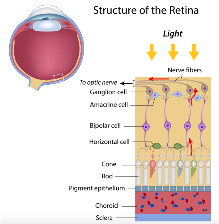
What are the two layers of the retina?
Pigmented Layer: retinal pigment epithelium. contains lots of melanin to ensure no excessive refraction of light rays and stop glare. (albinos don’t have melanin so this is why they struggle with normal light being too bright)
Neural Layer: contains photoreceptors (rods and cones) and horizontal cells which do lateral inhibition that stops neighbouring cells to the highest intensity cells from detecting the light. Bipolar cells are found between photoreceptors and ganglion cells
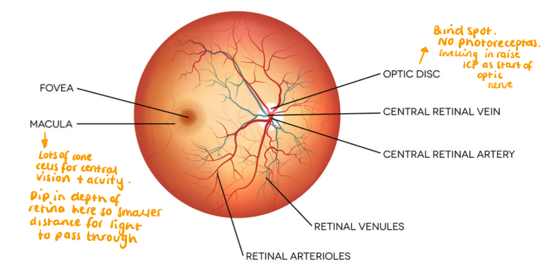
What is the blood supply to the retina?
- Comes from the choroid later from the central retinal artery
- Can get occluded due to atherosclerosis of the ICA
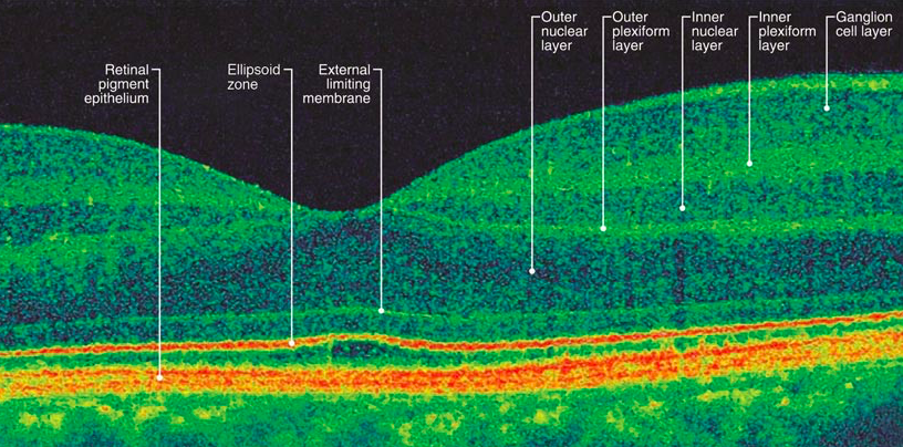
How can we image the retina?
Optical Coherence Tomography
What are some pathological processes that can affect the retina?
- Hypertension and diabetes can cause retinopathy
- Amaurosis Fugax in a stroke
- Macula degeneration
- Papilloedema in raised ICP
What is Amaurosis Fugax?
- Transient monocular blindness
- CURTAIN COMING DOWN OVER VISION - THINK STROKE
- Due to blockage in ICA or retinal artery

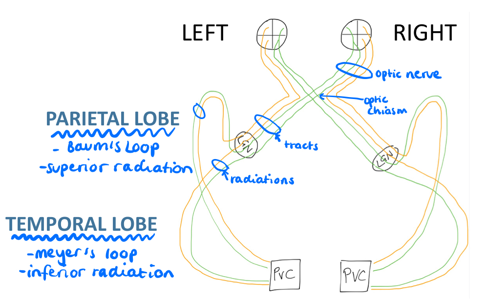
What are the components of the visual pathway and draw a diagram displaying the pathway?
Optic Nerve –> Optic Chiasm –> Optic Tracts –> Lateral Geniculate Nucleus in the Thalamus –> Radiations (superior and inferior) –> Occipital Lobe (primary visual cortex)
What fibres are in the left superior radiation (Baum’s loop)?
- Left superior temporal fibre and right superior nasal fibre
- If in upper quadrant, in superior radiation and vice versa with lower quadrant

Where are each of the radiations?
Superior: parietal lobe
Inferior: temporal lobe
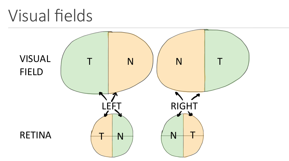
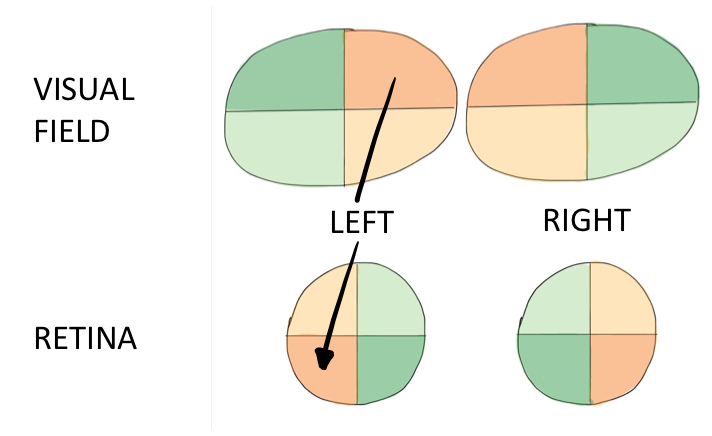
What fibres make up the optic nerve and what part of the visual field does each fibre contribute to?
- Nasal retinal fibres responsible for temporal visual field
- Temporal retinal fibres responsible for nasal visual field
- Due to the fact light travels in straight lines
- 4 fibres make up the nerve (superior and inferior)
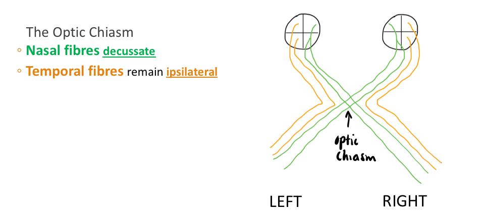
What is the difference between temporal and nasal fibres?
Where are the optic tracts?
After the optic chiasm up to the lateral geniculate nucleus
What is the best way to figure out where a lesion is in the visual pathway?
Visual field defects are names on area of visual loss not the lesion so draw the diagram out!!!
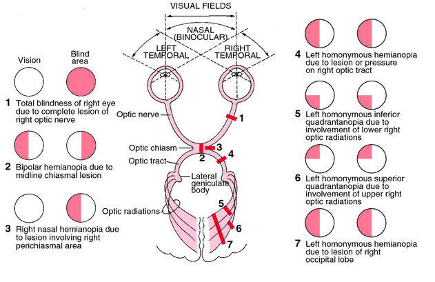
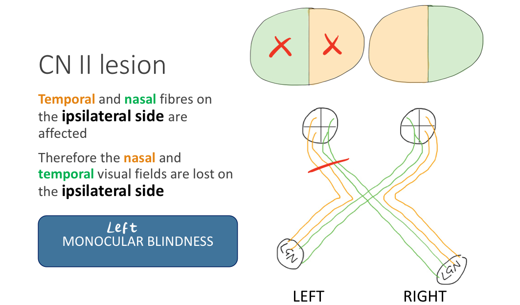
What is monocular blindness and where is the lesion in this case?
- Complete vision loss in on eye
- Can be due to retinoblastoma, meningiomas or blockage in central retinal artery (stroke)
- Lesion in optic nerve so ipsilateral temporal and nasal fibres lost
What type of visual field loss is this, and where is the lesion in the visual pathway?

Bitemporal Hemianopia
- Lesion in optic chiasm
- Loss of nasal fibres on both sides so you lose your temporal visual field, causing tunnel vision
- Pituitary gland tumour or anterior communicating artery aneurysm

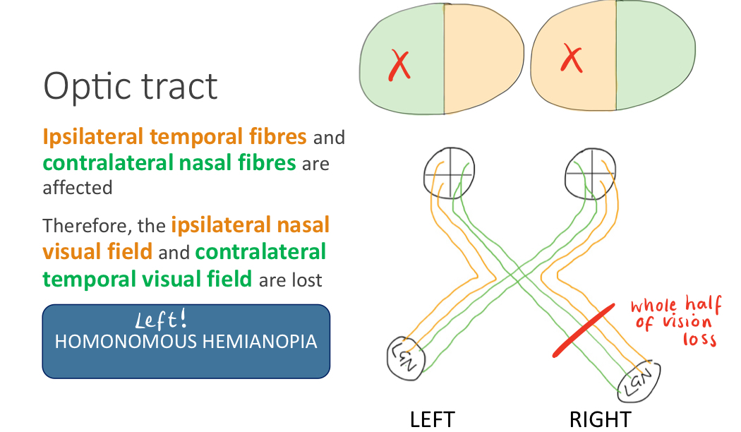
What type of visual field loss is this, and where is the lesion in the visual pathway?

Left homonymous hemianopia
- Lesion in the right optic tract. Contralateral lesion to vision loss, use nasal field loss to guide side of lesion as temporal fibres don’t decussate
- Could be neoplasia or trauma
What part of the visual field do the optic radiations supply?
- Inferior radiations supply superior visual vield and vice versa
What type of visual field loss is this and where is the lesion?

- Left homonymous inferior quadrantanopia
- Lesion in right superior optic radiation (parietal lobe)

What type of visual field loss is this and where is the lesion?

Right superior homonymous quandrantanopia
- Left inferior radiation lesion
Apart from a lesion in the optic tract, what other pathology could result in a homonymous hemianopia?
A stroke affecting the MCA could knock out both radiations ipsilaterally, have to look at the clinical history to distinguish between stroke and damage to a tract

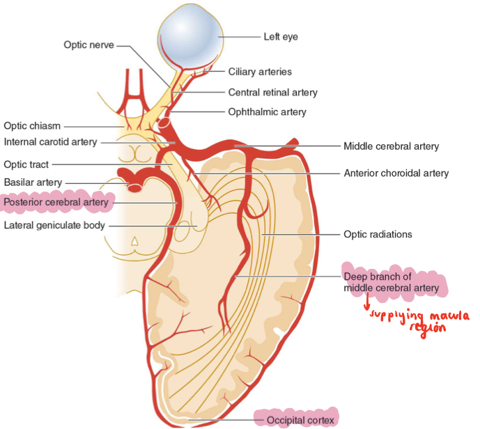
What are these visual field defects showing and why does this occur?

- Macular Sparing: still good central vision
- Occipital lobe has dual blood supply from PCA and MCA.
- If there is a stroke of the PCA, most of the occipital lobe is lost but the MCA supplies the occipital pole(macula) so vision is spared centally
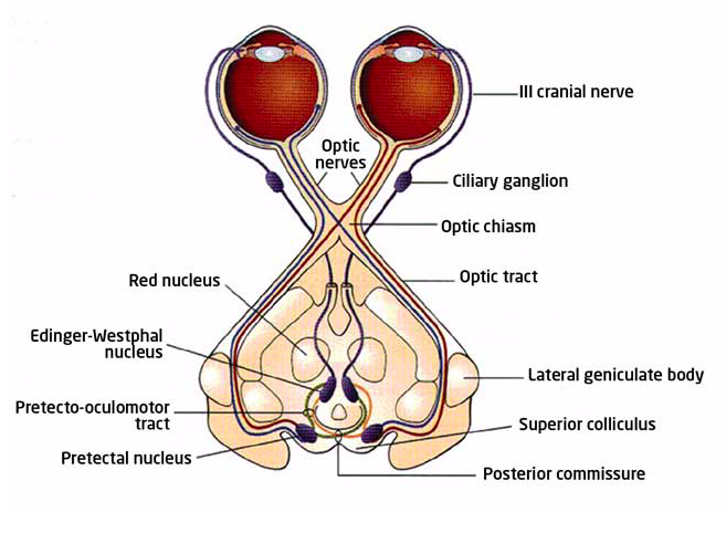
Draw the nerve pathway involved in the light reflex.
- Light stimulates CNII and goes on to synaps with Edinger Westphal nucleus in pre tectal area
- Parasympathetic respone through efferent CNIII
- Direct and consensual pupil constriction via constricter pupillae
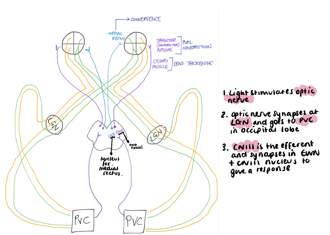
Draw the nerve pathway involved in the accomodation reflex.
3 C’s:
- Convergence (medial rectus)
- Constriction (constrictor pupillae)
- Convexivity so fat lens (cilliary muscles)
Cerebral cortex involved as well as midbrain as some image interpretation unlike in the light reflex. Occipital lobe sends fibres back up to midbrain
Label the cranial nerves on this cadaveric specimen.


Where does the fundus sit in relation to the optic disc?
Lateral


-
1 - Topography of the Nervous System30
-
2 - Embryology of the Nervous System18
-
3 - Glia36
-
4 - Somatosensory System32
-
5 - The Eye43
-
6 - Arterial Supply to the Brain17
-
7 - The Motor System40
-
8 - Movement Disorders46
-
9 - Higher Cortical Function22
-
10 - Reticular Formation and Consciousness24
-
11 - Neuropathology35
-
12 - Confusion in the Elderly36
-
13 - Headaches40
-
14 - Stroke27
-
15 - Raised ICP27
-
16 - Meningitis and Subarachnoid Haemorraghe34
-
17 - Anxiety Disorders24
-
18 - Mood Disorders28
-
19 - Psychosis31
