Neuropathology 2 Flashcards

(164 cards)

1
Q

A: Describe [Radicular Root Pain]

B: What causes it (2)

C: What, associated with this, causes a [dull & local pain]?

A

A: [LSS Pain-Lightning/Stabbing/Shooting Pain] that situates itself in the dermatomal distribution of a dorsal root

B:

  1. Inflammation of [Dorsal Root]
  2. Extramedullary compression of [Dorsal Root]

C: The extramedullary lesion itself

2
Q

A: Clinical sensory deficits correlate to the ____[vertebrae / spinal cord] level

B: Which between the two listed above extends down longer?

A

A: Clinical sensory deficits correlate to the Spinal Cord level

B: Vertebral column becomes longer than spinal cord during development

3
Q

Describe the sensory signs for Spinothalamic Tract lesions (3)

A
  1. Contralateral deficit of pain and temp
  2. [Sacral Sparing during intramedullary lesions] (since sacral fibers are far lateral)
  3. [Deficit is up to dermatomal level in EXTRAmedullary lesions]
4
Q

A: Clinical Manifestation for [ANT Spinal Artery Occlusion] (3)

B: What types of things cause this?

A

A:

1) Sudden Hyperreflexic spastic paraparesis
2) Loss of Pain/Temp inferior to the lesion
3) Preserved [2TVP-2point/Touch/Vibration/Position]

B:

  • Atherosclerotic aortic Dz
  • Aortic Surgery
5
Q

A: Describe [SuB ACute Combined Degeneration] (2)

B: What are the causes (3)

A

A: [Demyelinating lesions] in Posterior and Lateral Columns (usually at thoracic level) –>

  • loss of [2TVP- 2point discrimination/Touch/vibration/position] of LE
  • but with…*
  • intact Pain and Temp

B: [SuB ACute Combined Degeneration]

1) B12 Deficiency
2) Copper Deficiency
3) AIDS/HIV

6
Q

A: Describe [ALS-Amyotrophic Lateral Sclerosis]

B: Clinical Manifestation (3)

C: Prognosis and Tx

D: Issue with diagnosing ALS

E: Breathing Evaluation (2)

A

A: Progressive Degeneration of [UMN Pyrimidal Betz Cells] AND [LMN ANT Horn cells <–AFFECTED MORE!] –> Spheroid lesions

B:

1) Weakness affecting speech/chewing/breathing and eventually proximal limb atrophy = LMN sign
2) Fasciculations Diffusely = LMN sign
3) [Exaggerated Reflexes + Babinski] = UMN sign

C: Fatal but can give [Riluzole (glutamate blocker)] since glutamate over exites motor neurons

D: UMN signs may be first confused with a cervical spinal cord lesion!

E: many pts fear respiratory failure. Mitigate with:

  • Aggressive = [Tracheostomy Mechanical Ventilation]
  • Supportive= [CPAP vs. BiPAP]
7
Q

Motor Neuron Disease

A: ALS-Amyotrophic Lateral Sclerosis Pgn

B: Mode of Inheritance

A

A: Progressive Degeneration of [UMN Pyrimidal Betz Cells] AND [LMN ANT Horn cells <–AFFECTED MORE!] –> Spheroid lesions & has the WORST PGN OF ALL MOTOR NEURON DISORDERS (50% Die within 3 years from respiratory failure or profound weakness)

B: [RARELY FAMILIAL (Chromo 21 Superoxide Dismutase Gene mutation) but affects more Males]

8
Q

Clinical Manifestation of Tabes Dorsalis (4)

A

1st) Lightning pain from [initial dorsal root lesion] (from loss of DRG and dorsal root)
2nd) Loss of [2VP-2point discrimination/vibration/position] from dorsal column degeneration –> [Romberg] + [Stomping Gait] + [Charcot Joints]
3rd) Loss of ALL SENSES (from loss of DRG and dorsal root)
4th) [Areflexia but Preserved Strength]

9
Q

A: Describe the 3 main sx for [Brown Sequard Syndrome]

B: Causes (3)

A
  1. Contralateral STT Loss of Pain/Temp
  2. Ipsilateral DCP Loss of 2TVP-2point/Touch/Vibration/[Position Proprioreception]
    * 3.* Ipsilateral CST Loss –> Muscle Weakness

B:

[(Extramedullary Tumor] vs. Trauma vs. (Herniated Disc)]

10
Q

A: Pathophysiology of Myasthenia Gravis

B: When does it onset? Describe the 2 types.

C: What things are preserved in this dz? (2)

D: What is Myasthenic Fatigue

A

A: Autoimmune Dz that blocks and INC degradative turnover of [postsynpatic nicotinic ACh Receptors]]

B: [Generalized (more common) vs. Ocular] and occurs at ANY AGE!

  1. Generalized= P DDD WF

[Ptosis/[Diplopia from Disconjugate gaze]/Dysarthria/Dysphagia/ [Weakness(Respiratory and limbs)/ Fatigue-especially with certain activities] ]

  1. Ocular= [Ptosis/Diplopia] after 2-3 years of dx

C: Sensation and Reflexes

D: Exercise normally DEC ACh release but is compensated by the saftey factor.

During Myasthenia Gravis, this DEC ACh during exercise PLUS the [Loss of EPP-End Plate Potential]–> Loss of muscle depolarization –> Myasthenic Fatigue

11
Q

A: 3 ways to diagnose Myasthenia Gravis

B: Which test is most specific

A
  1. Elevated [Serum Antibody against (postsynpatic nicotinic ACh Receptors)] = MOST SPECIFIC
  2. Positive [Tensilon Edrophonium] : short acting AChEsterase inhibitor. If after IV injection, pt feels better = they have Myasthenia Gravis
  3. EMG showing evidence of abnormal Neuromuscular junction transmission (i.e. repetitive nerve stimulation)
12
Q

Myasthenia Gravis Tx (4)

A
  1. AntiCholinesterase drugs –> allows ACh to stick around longer
  2. Thymectomy (remove part of Thymus that contains ACh Receptor-like material)
  3. Immunosuppresants

  • -[Azthioprine vs. Mycophenolate Mofetil]*
  • -Cyclosporine*
  • -Corticosteroids*
    4. [Plasmapheresis vs. IV Immunoglobulin]= Transient but potent fixes
13
Q

A: Pathophysiology of [Lambert Eaton Myasthenic Syndrome]

B: Clinical Manifestation (3)

C: Dx (3)

D: What CA is this syndrome associated with?

A

A: [Autoimmune attack against (Presynpatic Ca+ channel)–> No ACh release]

B:

  • Fatigable weakness of Proximal limbs and trunk that mimic myopathy
  • Improved briefly by exertion
  • Autonomic sx (Dry mouth & Orthostasis)

C: [Nerve Stimulation test vs. EMG vs. Ab Detection]

D: usually associated with SOLC-Small Oat cell Lung Carcinoma

14
Q

[Lambert Eaton Myasthenic Syndrome] tx (3)

A
  1. Tx underlying CA
  2. Drugs to enhance ACh release (Guanidine vs. Diaminopyridine)
  3. Immunosuppresants
15
Q

A: What is Mononeuropathy

B: Pathophysiology

C: Dx (2)

D: Examples (3)

A

A: Single Major “named” nerve is involved (sensory vs. motor vs. Both)

B: [Trauma or Compression] —> focal demyelination of a nerve and possibly axonal damage if lesion is severe

C: Diagnosed with EMG and nerve testing

D: Ex:

  1. [Carpal Tunnel Median mononeuropathy] = most common!
  2. Ulnar mononeuropathy from leaning on elbow
  3. Peroneal mononeropathy from [lateral knee injury]
16
Q

A: [Carpal Tunnel Median mononeuropathy] pathophysiology

A2: What do severe cases of this lead to?

B: Tx (3)

A

A: is a compression mononeuropathy that occurs when [inflammed flexor tendons/fluid retention (pregnancy) / swelling] all compress the Median nerve in the carpal tunnel –> Tingling Numbness.

A2: Severe cases = Thenar atrophy–> weakness

B: Tx

  • Anti-Inflammatory
  • Local Rest
  • Surgery vs. Splint
  • MOST COMMON MONONEUROPATHY*
17
Q

What is Wallerian Degeneration?

A

When the peri and epineurium are preserved after the nerve trauma the axons undergoes Wallerian Degeneration. The perserved scaffolding allows sufficient axonal sprouting and regeneration within the PNS

18
Q

A: Pathophysiology of [Peripheral Polyneuropathy]

B: Clinical Manifestation of [Peripheral Polyneuropathy] (4)

A

A: [Disorder of multiple, major AND small n.] caused by -axonal degeneration(will DEC EMG amplitude) and -secondary demyelination(will DEC EMG velocity). Both due to inadequate axoplasmic flow–>

B: 1. [early sensory loss of distal limbs (i.e. feet)]–> eventually [motor loss of distal limbs] –> atrophy–> weakness. (longest sensory cells are affected 1st)

  1. [Early loss of muscle stretch reflexes]
  2. Paresthesia= spontaneous tingling
  3. Dysesthesia= Unpleasant sensation from non-noxious stimulus
    * eventually hands are affected as well*
19
Q

A: Causes of [Peripheral Polyneuropathy] (4)

B: Dx (3)

A
  1. Hereditary
  2. Toxic (Drugs vs. Occupation)
  3. Other Multiple Mononeuropathies or autoimmune pathologies (DM vs. SLE vs. Guillain Barre)
  4. Idiopathic= MOST COMMON

B: EMG vs Blood testing vs. Nerve biopsy

20
Q

Describe the EMG

A

Needle Electromyography. Electrical activity of muscles within 1 motor unit to be assessed for nerve damage and muscle dz

21
Q

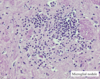
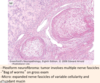
A: Describe Guillain Barre Syndrome

B: What Pt demographic is mostly affected by this

A

A: Acute Polyneuropathy manifesting as inflammation and demyelination of [peripheral n. and roots] –> [ascending NON-reflexic paralysis (includes respiratory paralysis)] –> [little sensory loss but some paresthesia and eventually reflex loss]

B: Occurs at any age, but 50% of pts have [Viral URI] prior to getting Guillain Barre

22
Q

Guillain Barre Syndrome

A: Dx (2)

B: Pgn

C: What tx would help accelerate recovery?

A

A: Dx

1) EMG testing that reveals demyelination
2) Elevated CSF Protein and possible WBC

B: Pgn = GOOD!

C: [Plasmapheresis vs. IVIG] may shorten illness

Remember that Guillain Barre is a type of Polyneuropathy

23
Q

[Chronic Acquired Polyneuropathies]

Causes (8)

A

[Chronic Acquired Polyneuropathies] : takes months-years to actually develop

May Destroy ​NITRIC

  1. Metabolic/Endocrine (Uremia vs. hypOthyroid)
  2. DM
  3. Nutrition (Vitamin B Deficiency)
  4. Infection (Leprosy = MOST COMMON WORLDWIDE)
  5. Toxins (alcoholism vs. lead)
  6. Rheumatological (RA vs. Lupus)
  7. Idiopathic
  8. CA (myeloma)
24
Q

Hereditary Neuropathies

A: Pathophysiology

B: When does it onset

C: Clinical Manifestation and tx

A

A: known or unknown metabolic vs. genetic disorders –> [Distal sensorimotor deficits with little to no paresthesia/dysesthesia]

B: Childhood

C: Orthopedic Deformities (scoliosis/hammertoes/pes cavus): Give assistive devices, but otherwise NO TX

25
A: **Myopathy** Clinical Manifestation (3) B: Dx (4)
A: 1) **Proximal** Limb (*shoulders/hips*) weakness and atrophy 2) *LATE* loss of reflexes after muscular atrophy onsets 3) Intact sensation B: - Family hx of muscualar dystrophy - Elevated Creatinine Kinase (*muscle enzyme*) - EMG - Muscle Biopsy
26
A: **Polymyositis** (Describe and List causes (4)) B: Clinical Manifestation (2) C: Dx D: Tx
A: A type of *myopathy* involving **multiple** muscles and caused by: 1) Autoimmune= MOST COMMON IN USA 2) Viral 3) Drugs 4) CA= rare B: - Proximal weakness over weeks to months - Dermatomyositis: *Rash around eyes or fingers* C: \*[Inflammation-mononuclear inflammatory infiltration]/necrosis on biopsy D: Corticosteroids
27
**Duchenne's Muscular Dystrophy** A: pathophysiology B: Onset C: Clinical Manifestation (2)
A: [**X-linked disorder**] --\> Absence of the [dystrophin muscular structural protein]--\> [Cardiac / Respiratory / *Proximal* Limb Weakness] B: **Boy**hood C: 1) Calf Pseudohypertrophy (*from muscle being replaced by fat & connective tissue*) 2) Cardiorespiratory Death by Age 30
28
A: Describe **Motor Neuron Disease** B: MOD for sub-type: ***Spinal Muscular Atrophy*** - Clinical Manifestation - **Infant** type: Name and MOD - **Teen/Adult** type: Pgn
A: Degeneration of **UMN**, **LMN**, **or both** --\> varying serverity and rate of degeneration B: _[Spinal muscular atrophy]_ = specifically [ANT Horn Cell degeneration] from [*Chromo 5* SMN1 and 2 gene mutations]--\> LMN signs of FAW- ***W**eakness/[**a**trophy & areflexia] /**F**asciculations* *\**Infantile onset = (**Werdnig Hoffman**) --\> [Auto**R**ecessive terminal condition --\> *Floppy Baby* from defuse [Distal muscle atrophy] \*Milder childhood/adult onset types --\> [Non-fatal Chronic Disability]
29
A: Clinical Manifestation of **Olfactory Nerve** lesion (3) B: How are these lesions associated with *Head Trauma*?
A: 1) [Sinusitis vs. URI] (**most common**) 2) Anosmia 3) Orbitofrontal Tumor (**rare**) B: Shears nerve branches in *Cribiform Plate*
30
A: Describe the difference between **Binocular Diplopia** and **monocular Diplopia** B: Lesion of [CN 3/4/6] produces what type of Diplopia?
A: Binocular= single image appears when 1 eye is covered vs. monocular= **DOUBLE** image appears when 1 eye is covered (*comes from Psych dz vs. [ocular pathology i.e. dislocated lens]*) B: Binocular Diplopia
31
A: Clinical Manifestation of **Pineal Tumors** B: Mechanism of Dz C: *Other* Causes with _same manifestation_ (2)
A: Pupil constricts with near reflex **but not with light** B: Selective disruption of [light reflex pathway] in [**Pretectal Midbrain**] C: 1) Neurosyphilis (*Argyll Robertson pupil*) 2) [Dorsal midbrain lesions] (*Pineal tumors-* *Parinaud's Syndrome* - *will also have poor upgaze*
32
A: What is [**MLF**-Medial Longitudinal Fasciculus] syndrome AKA? B: Clinical Presentation (3) C: What causes **MLF syndrome** in younger vs. Older pts?
A: Internuclear Ophthalmoplegia [**MIOS**-***M**LF **I**nternuclear **O**phthalmoplegia **S**yndrome*] B: \*[Impaired **ADD**uction of **affected** eye] + [Normal **ADD**uction of **affected** eye *during [near reflex convergence]* + \*[Nystagmus of **UN**affected eye when attempting to A*B*duct] C: 1) Younger pts= Multiple Sclerosis 2) Older pts= Ischemic infarction
33
What are the 4 common HA
1. **Tension** 2. **Migraines** 3. **[Fever/Hunger provoked]** 4. **HEENT related**
34
*HA Red Flags:* What **[signs & sx concomitant with Acute onset HA]** make you suspect [Aneurysmal SubArachnoid Hemorrhage] _or_ [Cerebellar Hematoma] (5)
1. Split second / unexpected 2. Worst or not previously encountered 3. Loss of Consciousness 4. Vertigo 5. Vomiting
35
*HA Red Flags:* [**Fever & Skin Rash**] with *Acute Onset HA* may indicate what *HA Dx*?
Meningitis ## Footnote (*keep in mind that Acute Meningitis evolves _rapidly_, including impaired consciousness / nuchal rigitidity / NV*)
36
*HA Red Flags:* [**Immunocompromised pts**] with *Acute Onset HA* may indicate what *HA Dx*? (2)
1. CryptoSporidium *Meningitis* 2. Toxoplasmosis
37
*HA Red Flags:* [**Coagulopathy / anticoagulation**] with *Acute Onset HA* may indicate what *HA Dx*?
[SubDural vs. intradural **Hematoma**]
38
A: What are the *3 Clinical Questions* that help to diagnose **Migraine**? B: **Migraine** *General Characteristics* (6)
A: 1) Have *nausea* when u have HA? 2) HA exacerbated with *light*? 3) HA limit you from *everyday functioning*? B: "**DUCAP** gives me *Migraines*, and *Migraines* gives me PANTOS​" **P**eriodic **A**ctivity-limiting [**D**iminishes in freq. throughout life] (*Rare for older pts to have migraines for 1st time*) **U**nilateral & Pulsating usually **C**hildhood-*late* vs. early adult onset
39
Name 5 **Migraine** triggers
1. Stress 2. Sleep Deprivation 3. Hunger vs. Foods 4. Alcohol/nitrates 5. Fumes/smoke
40
***Migraine** Phases:* **Prodrome** A: Duration B: Sx (6)
A: **Pro**drome: [6-48 hour duration] occuring **BEFORE Migraine** **R**ight **I**n **F**ront of **DD**angerous **H**A [**R**hinorrhea vs. Lacrimation] **I**rratilibility **F**atigue **D**epression *and* **D**rowsiness **H**unger (Cravings for chocolate/nuts/bananas)
41
***Migraine** Phases:* **Aura** A: Duration B: Define *Acephalgic Migraine* C: Describe **Aura**
A: **Aura**: [Before \>during\>\> after] **Migraine**. [Migraine HA] onsets within an hour after experiencing Aura, but Aura last *no longer* than 1 hour. B: _Aura May not be present with Migraine_. = Acephalgic Migraine C: [Visual sx (most common)= blind spot near center of vision w/flashing & pulsating bands of light spreading across + diplopia
42
***Migraine** Phases:* **PAIN** A: Location (3) B: Duration C: Associated sx (7)
A: **Pain:** [Head \> Abd \> Precordial] B: _[(_minutes to hour **gradual** onset) with (Hours to Days duration)] C: "DUCAP gives me *Migraines*, and *Migraines* gives me **PANTOS**" - [**P**ho*T*ophobia vs. Pho*N*ophobia] - [**A**phasia + Dizziness (*Cortex involvement*)] - **N**/V - **T**hermophobia - **O**smophobia (*fear of odors*) - **S**ensorimotor deficit (*Tingling*)
43
**Migraine** etx ; How are the*Trigeminal nerves* associated-2
Genetic [GainOfFunction mutation in *excitatory NMDA receptor*]--\>burst of cerebral activity _when triggered_---\>hyperemia (*usually occipital lobe*)--\> sx. Burst is followed by ⬇︎ cortical activity= **Cortical Depression** tht has slow but deliberate forward advance --\> Triggers Trigeminal pathway Trigeminal afferents : 1. send impulses--\>Brain Stem & hypothalamus--\> Nausea/Photophobia/Phonophobia 2. retroactively depolarize--\>release of substance P --\> neurogenic inflammatory pain + vasoDilation
44
***Migraine** Phases:* **PostDrome** A: Duration B: Sx (3)
A: **PostDrome:** [last several hours after] B: - Mood change (euphoria vs. fatigue) - concentration problems - Scalp/muscle tenderness
45
**Migraine** A: Tx (5) B: 3 things to keep in mind when treating **Migraine** C: *Alternative* Tx (5)
A: [*Effective Dose* **NSAID**] \> Triptans \> [Butalbital (*habit forming*) \> DHE \> Ergotamine B: - Treat **EARLY** (including Associated sx)! - Less is Best! - Px should be used when attacks\> 2-3/month, severe and abortive therapies have failed C: Botox vs. Butterbur vs. [St.John's Wort] vs. Ginger vs. VitB2
46
**Triptans Rx** A: MOA B: When are these indicated (2) C: When is the pt relieved of pain?
A: [5HT1B/D Receptor **Agonist**] --\> vaso**constriction**--\> relieves Migraines B: 1. Pt fails to respond to [*Effective Dose* **NSAID**] 2. Pt has Moderate-to-Severe Migraine Pain C: 2 (*more common)* vs. 24 Hours
47
**Triptans Rx** A: *Cx (6)* B: *Side Effects (2)* - Description and location - Duration
A: Contraindications: 1. Vascular Risk Factors (*Ischemic Heart Dz / uncontrolled HTN / Renal Dz*) 2. Pregnancy 3. [Basilar or Hemiplegic Migraine] 4. Avoid within 24 hours of Ergotamine 5. Pts taking [MAOI] 6. Renal or Hepatic impairment (**Ergotamine Cx**) 7. Age \> 60 (**Ergotamine Cx**​) B: Side Effects = 1. ***Triptan Sensations*** - [Pressure/Pain/Tightness/Warmth/Tingling] commonly in [Face/Limbs/Chest] but can occur anywhere \*\*Short duration and resolve spontaneously + 2. Serotonin Syndrome (*excessive 5HT**1a and 2** receptor activation*)--\> [Dysautonomia (Diarrhea / lacrimation)] + Encephalopathy
48
A: **Ergotamine** - Route of Administration - SE (2) B: **DHE** -Route of Administration (3)
*Migraine specific Rx* ## Footnote A: **Ergotamine (arterial vasoconstrictor)** - Route of Administration = Suppository ONLY - [Severe Nausea] vs. Uterine Contractions B: **DHE (arterial & venous vasoconstrictor)** -Route of Administration = [IV vs. SubQ vs. Nasal Spray]
49
A: **Botulinum Toxin** Indication B: Onset & Duration
A: [**Chronic Migraine** HA] AFTER pt has failed other meds (*works by relaxing muscles*) B: Takes a week to onset but will last up to 12 weeks
50
A: **Cluster HA** Dx Criteria B: location C: Demographic D: Tx (5)
A: **Rapid onset (****15-30 min)** of([1-4 Attacks/day] each lasting [20 min - 3 Hr] x [6-12 weeks] B: **ALWAYS UNILATERAL** C: [Men during spring & fall] D: - Inhaled O2 100% - *Injectable* Sumatriptan vs. Nasal Triptan - Nasal Lidocaine - Nasal DHE - Prednisone
51
A: Describe **Tension HA** B: Associated sx C: Clinical Presentation D: *Common* Causes (3)
A: [Classic Everyday ***Bilateral*** HA]= Most common HA B: Pho**N**ophobia _vs._ Pho**T**ophobia (*NOT AT SAME TIME*) C: [Pressing or Tightening Band head pain] lasting [4-6 Hr] maybe brought about by: - TCA rx - sleep deprivation - depression
52
A: Describe **Trigeminal Neuralgia** B: Tx C: *Most common* Cause
A: SEVERE Paroxysmal attacks of jaw-radiating pain lasting [ B: [**Carbamazepine** **200-1200 mg/day**] C: [Young people with Multiple Sclerosis]
53
A: Describe **Pseudotumor Cerebri** B: Demographic C: Clinical Presentation (2) D: Tx (3)
A: HA that can make *(mostly young Female)* pts go **Blind**! B: Overwt young female taking BCP C: Papilledema + [High ICP(*\>250*) confirmed with spinal tap] D: Topiramate(*will also --\> Wt loss :-)* ) vs. Acetazolamide vs. Surgery
54
A: Clinical Presentation of **Multiple Sclerosis** (3) B: Demographic C: *Associated* Causes (4)
A: "Charcot had *MS* and thought it was a **SIN**" **Charcot's Triad** 1. **N**ystagmus 2. [**I**ntention Tremor] 3. [**S**canning Speech] B: *Mostly* [Young White Female] but can be anyone especially those with [low Vitamin D/sun exposure] C: 1. Genetic *Contribution (HLA & SNP)* but MS is **not Genetic Dz** 2. Lack of Sun --\> low Vitamin D 3. Viral: *EBV / [Canine Distemper Virus]* 4. Tobacco
55
A: **Multiple Sclerosis** MOD B: Clinical Course (4)
A: Activated [Autoreactive T Cells] travel from peripheral lymph node to CNS--\>break down BBB w/interleukins that induce inflammation---\> then secrete inflammatory cytokines --\> [myelin destruction and neuronal death of **White** mater] B: [Sx\>24 hrs from demyelination]--\> [remission *(complete vs. partial ​improvmnt)*] --\> [Relapses every 1-2 years x 5-10 years] --\> [Progressive sx with no more relapses/new lesions]
56
Name the 5 *most common _syndromes_* associated with **Multile Sclerosis**
* Optic Neuritis * Afferent Pupillary Defect * [Brainstem Syndromes (***Internuclear Ophthalmoplegia / Ataxia / Trigeminal Neuralgia***) * [Spinal Cord Syndromes] * Romberg (*MS lesions "love" dorsal column destruction*)
57
*Optic Neuritis* is a syndrome highly associated with **Multiple Sclerosis** B: Sx (3) C: Other phenomena *Opitc Neuritis* my be associated with (2)
***Unilateral** Optic Disc Swelling on fundoscopic* ## Footnote 1. [Scotoma Blindspot] vs. [Complete Blindness] 2. Eye mvmnt pain 3. DEC [Red/Green] C: - [Marcus Gunn *Afferent* Pupil defect] - Uhthoff Phenomenon (*heat intolerance after being placed in hot tube*)
58
*Brainstem Syndromes* is a syndrome highly associated with **MS** B: What's the biggest example
B: [**MIOS-*****M**LF**I**nternuclear**O**phthalmoplegia**S**yndrome*]
59
*Spinal Cord Syndromes* is a syndrome highly associated with **MS** B: Classic *signs* (4) C: How do you differentiate this from **Guillan Barre** D: Description of location of spinal cord **MS** lesions
*Pts w/"Sensory Level" should be assumed to have Spinal Cord Lesion until proven otherwise. Not MS* ## Footnote B: - [Lhermitte's Sign]: Pain when chin is touched to chest - Deafferented Hand - Urinary sx + Erectile Dysfunction - Progressive **A**symmetric spastic paraplegia C: [Guillain Barre Pts] do **NOT** get numb in torso D: small, located in cord periphery (especially in dorsal columns). Begin at pial surface.
60
A: Dx Criteria for **Multiple Sclerosis** (4) B: Location of **MS** lesions (5) C: Which location lesion is shown in image? D: Which Radiography is used for dx? (2)
*Dx of MS is a clinical dx + radiographic corroboration* ## Footnote A: - [2 or more separate parts of CNS involved] - [2 or more worsening episodes (*separated by 1 month or more and each lasting 24 hrs*)] - Radiography: [Periventricular **White** Matter lesion (*\> 3mm*) disseminated in time & space] = Dx of Exclusion - CSF Fluid with [Oligoclonal bands] + [DEC Cell count] + [IgG abnormalitites] + [Normal Glucose & Protein] = **NOT MANDATORY** B: [Corpus Callosum] / [Optic Radiation] / [Brainstem abutting 4th vt] / [T1 = Black Holes] / [**Dawson's Finger on T2W (shown in image)**] C: [**Dawson's Finger on T2W (shown in image)**] D: [MRI: T**2**W vs. FLAIR] (*will have **H**yperdense/**H**igh Signal)*
61
Common other DDx of [**MS**​-like presentation] include [Neuromyelitis Optica] vs. [ADEM] vs. [PML] ## Footnote * Describe **Neuromyelitis Optica*** * B: Clinical Presentation (3)*
**Longitudinally extensive** spinal cord lesion (*\>3 vertebrae*) --\> Bilateral optic neuritis ## Footnote B: - Hic-coughs - Normal Brain - [NMO IgG Ab Positive] - aquaporin 4 channel
62
Common other DDx of [**MS**​-like presentation] include [Neuromyelitis Optica] vs. [ADEM] vs. [PML] ## Footnote * Describe [**ADEM-A**cute **D**isseminated **E**ncephalo**M**yelitis]* * A: Onset* * B: Associated sx (3)* * C: Demographic*
A: Post-meningoencephalitis/infectious [Large *FLUFFY Multifocal] lesions* B: - HA - Vomiting - Drowsiness - Meningism C: Children
63
Common other DDx of [**MS**-like presentation] include [Neuromyelitis Optica] vs. [ADEM] vs. [PML] ## Footnote * Describe **PML-P**rogressive **M**ultifocal **L**eukoencephalopathy* * B: Location (2)* * C: How is **PML** related to the drug, Natalizumab?* * D: Demographic* * E: Histology (3)*
Opportunistic infection 2º to [John Cunningham PolyomaVirus]----\> [**multiple white** matter lesions] (***Hyperintense Flair signal on radiology***) --\> Death vs. Severe Neuro injury ## Footnote B: [SubCortical Hemispheric White Matter] or [Cerebellar Peduncles] C: Also can be caused by Rare Side Effect of **Natalizumab** (**MS** drug) in pts who are also JC Virus positive D: HIV pts (*reversal of immunosuppresion stops viral progression*) E: "PML's HISTO is like a **MOB**" - **M**yelin Loss (*with axonal sparing*) - **B**izarre astrocytes - **O**ligodendroglial inclusions (--\> *focal* *discoloration in white matter & Ground glass appearance*)
64
A: **Multiple Sclerosis** Tx (4) B: Which one is used for [Acute **MS** Relapses]?
A: Tx 1. [IV vs. Oral **Methylprednisolone**] (*IV is preferred*) 2. ACTH 3. Plasmapharesis 4. IVIG B: [IV vs. Oral **Methylprednisolone**] (*IV is preferred*)
65
Name the 5 major causes of **Meningitis** and descriptions of each
* Bacterial= Acute * Fungal= affects Immunocompromised * Amebic(*Parasitic*) & TB= Granulomatous * Viral= [**ASEPTIC** meningitis] & [self-limiting] * Non-Infectious= Chemical vs. [Meningeal Carcinomatosis]
66
**Acute Bacterial Meningitis** What common bacteria affect ages... A: Neonates B: 1-12 months (2) C: 1 - 16 years (3) D: 16 - 50 years (2) E: *Age Extremes (2)*
A: Neonates:- [E.Coli] vs. [Group B Strep] B: 1-12 months: [Strep Pneumo] vs. [H.Flu] C: 1 - 16 years: [Neisseria meningitis] vs. [H.Flu] vs. [Strep Pneumo] D: 16 - 50 years:[Neisseria meningitis] vs. [Strep Pneumo] E: *Age Extremes:* *[Listeria Monocytogenes] vs. [Pseudomonas Aeruginosa]*
67
**Describe CSF findings for *Bacteria* Meningitis** A: Fluid Quality B: Cells Present C: Protein level D: Glucose (*relative to plasma*) E: Pressure F: Tx if ***Bacteria* Meningitis** is suspected (2)
A: Cloudy B: PMNs C: **VERY HIGH PROTEIN** D: **Low Glucose** E: **HIGH PRESSURE** F: 1st: [**Emergent** IV Dexamethasone (*in adults*) + Broad Spectrum Abx] -----\> [specific abx after identification]
68
**Describe CSF findings for *Viral* Meningitis** A: Fluid Quality B: Cells Present C: Protein level D: Glucose (*relative to plasma*) E: Viral Causes (2)
A: Clear B: [Lymphocytes with lymphocytic extension along Virchow-Robin spaces] C: Slightly High Protein D: Normal Glucose E: 1) Enterovirus = Most Common 2) Arbovirus (*West Nile*)
69
**Describe CSF findings for *TB* Meningitis** A: Fluid Quality B: Cells Present C: Protein level D: Glucose (*relative to plasma*)
A: N/A B: Lymphocytes C: Moderately High Protein D: Mildly Low Glucose
70
List the 4 causes of **_Chronic_ Meningitis** *list examples* B: Demographics (3) C: Presentation (2)
*Evolves over weeks to months* 1. TB--\> necrotizing Granuloma (*PCR testing needed*) 2. Fungal (*Cryptococcus neoformans vs. Histoplasma vs. Coccidioides immitis*) 3. Parasitic (*Treponema Pallidum vs. Borrelia Burgdorferi*) = RARE 4. Non-Infectious (*Neurosarcoid*) B: Elderly vs. Malnourished vs. Immunosuppresed C: *Subtle* sx of HA and/or confusion with _no obvious meningeal signs_
71
In addition to Fungal **Meningitis**, fungus can also cause **\_\_\_\_\_\_,** **\_\_\_\_\_\_\_** and **[secondary vasculitis]** B: Clinical Manifestation of **[secondary vasculitis]** (2) C: Microscopy D: Stains used for dx (3)
In addition to Fungal **Meningitis**, fungus can also cause **encephalitis,** **Brain Abscess** and **[secondary vasculitis]** ## Footnote B: 1) Vascular invasion--\> infarct 2) Mycotic aneurysm--\> Hemorrhage C: Granulomatous Mononuclear infiltrate (*overlaps with TB*) D: [PAS vs. Mucicarmine vs. GMS]
72
**Fungal Meningitis** A: Which are *Hyphal & Pseudohyphal (4)* B: Which are *Yeast* *(3)*
A: **CAZF** [**C**andida/**A**spergillus/**Z**ygomycetes/**F**usarium] B: [Histoplasma/Blastomyces/Cryptococcus]
73
**Cysticercosis** / **Toxoplasmosis / Amoebiasis** all can cause **_Parasitic_ CNS infections** A: Describe **Cysticercosis** B: Geography (2)
MOST COMMON CEREBRAL PARASITE acquired via [Taenia solium Pork cestode] B: [SW states] and Mexico
74
*Taenia Solium Pork Cestode*
75
**Cysticercosis** / **Toxoplasmosis / Amoebiasis** all can cause **_Parasitic_ CNS infections** A: Describe **Toxoplasmosis** B: Acquisition (2) C: Radiology D: *Congential Toxoplasmosis* is part of \_\_\_\_\_ E: Dx (2)
Protozoan that crosses placenta (***pregnant women should avoid changing cat litter****)* B: Cysts in meat or [cat feces oocyst] C: Brain Abscess (***MRI Ring Enhancing Lesion***) D: -Congenital Toxo is part of **TORCH** (**T**oxo/**O**thers/**R**ubella/**C**MV/**H**erpes) D2: Dx = Serology vs. Biopsy
76
**Cysticercosis** / **Toxoplasmosis / Amoebiasis** all can cause **_Amoebiasis_ CNS infections** What 3 organisms are associated with Amoebiasis?
1. [Naegleria Fowleri Free living Amoeba] --\> [Fulminant Acute Meningoencephalitis] * Image shows _Trophozoites_ of Naegleria Fowleri* 2. Entamoeba Histolytica 3. Balamuthia Mandrillaris
77
**Viral _Encephalitis_** A: Major pathogens of [Summer/Early Fall] (3) B: [Fall and Winter] C: [Winter and Spring] D: [Any Season] (5)
A: Summer/Early Fall = **Arboviruses (West Nile Virus)** vs. Enterovirus vs. Rocky Mountain(*mimics viral encephalitis*) B: [Fall and Winter] = **LCMV** (**L**ymphocytic **C**horiomeningitis **V**irus) C: [Winter and Spring] = Mumps D: [Any Season] = **HSV1**/ EBV / CMV / Mycoplasma / Leptospira
78
**Viral _Encephalitis_** A: Affects White or Grey Matter? B: Histology (4)
A: Affects more **Grey** Matter (diffuse vs. focal) B: 1) [Perivascular Lymphocytic inflammation] 2) [Leptomeningeal Lymphocytic inflammation] 3) [Microglial cluster nodules] 4) Neuronophagia
79
A: **Polio Encephalitis** MOD B: Dx C: Which pathogen can this be confused with? D: Clinical Presentation
A: [**Fecal-Oral transmitted**] virus that replicates in [oropharynx & small intestine] & ultimately--\> [*ANT horn LMN* destruction in brainstem/spinal cord] B: Recovered from stool or throat C: Can mimic [**West Nile *Arbo*Virus**] & vice versa D: [*Asymmetrical* LMN signs = *Asymmetrical* **FAW** = **F**asciculations/[**A**trophy & areflexia] / **W**eakness]
80
A: Describe MOD for **Rabies** and what virus causes it B: Incubation period?
*Rhabdovirus* ## Footnote A: **Rabies**= Exposure to rapid dogs (*also bat/raccoon/skunk*) ---\> [Prodrome of flu-like sx] + [**Negri body cytoplasmic inclusions**] seen in brainstem/hippocampus/[Cerebellar Purkinje cells] B: [10 days to a year] depending on bite location
81
A: **Herpes Encephalitis** MOD B: Where does this Virus remain Latent? C: Histology D: What does the [*Burnt Out Herpes Encephalitis*] refer to? E: Tx
*Most commonly recognized and devastating* ## Footnote A: HSV1 (*transmitted via saliva*) --\> [(**Hemorrhage**---\>Cavitation & Atrophy) / Acute Necrosis/ Edema] of [**Medial Temporal & Frontal lobe]** B: Trigemial ganglion C: [Owl's Eye Intranuclear inclusion] D: _Chronic Phase_= Cavitation and Atrophy with shrivelled/brown color in long term survivors E: Acyclovir (*should be started even if suspected*)
82
A: **CMV** MOD B: Demographic (2) B2: Describe the Clinical Manifestation for each Demographic C: General Microscopy (2)
A: Opportunistic Virus (*Especially in AIDS and neonate pts*) that is is part of **TORCH** (**T**oxo/**O**thers/**R**ubella/**C**MV/**H**erpes) B: AIDS and neonate pts B1) Adults = Dilated CSF Ventricles and calcifications within periventricular region B2) Post**natal** infection = [multiple microglial nodules] + [occasional cytomegalic cellular inclusions] C: - Meningoencephalitis - [Cytomegalic cellular inclusions] mostly in periventricular regions
83
*[loss of myelin] and Atypical Astrocytes consistent with* ***PML**-**P**rogressive **M**ultifocal **L**eukoencephalopathy*
84
***HIVE*** **HIV Encephalopathy** B: Describe 2 other *neuro* conditions associated with HIV C: What Dx should you suspect in a *Young HIV Pt witih Dementia*? Pgn?
Widespread [microglial nodule **GREY MATTER ENCEPHALITIS**] ---\> [multinucleated giant cells] \*Also causes (*Meningitis--\>Persistent Pleocytosis & neuro sx)* and *Dementia* via _Direct_ Viral invasion vs. _inDirect_ inflammation C: **AIDS Dementia**= slow cognitive & behavioral decline with poor pgn. ***Note:** This presentation is Similar to [SubAcute Combined Degeneration]* *HIV LeukoEncephalopathy is the same thing but with White matter instead*
85
**HIV LeukoEncephalopathy** A: clinical presentation B: Histology C: Which brain cells are *NOT* affected by this?
A: Subacute onset with cognitive impairment and apathy B: [Diffuse **WHITE MATTER** myelin pallor with microglial nodules and (multinucleated giant cells) C: Oligodendrocytes are **NOT** infected *Unknown Etiology*
86
**Vacuolar Myelopathy**
## Footnote Spastic Paraparesis with hyperreflexia and ataxia caused by vacuolation of [Spinal Cord **White** Matter]
87
**Brain Abscess** A: Solitary or Multifocal B: Cause and [Dx method (3)] C: Tx (2) D: How are **Brain Abscess** related to CA
A: Usually Solitary (*can be multifocal*) within epidural or subdural of brain/spinal cord--\> 20% Mortality B: Caused by CNS infection that has to be indentified with [biopsy vs. aspiration vs. CT/MRI]. (***LUMBAR PUNCTURE MAY CAUSE HERNIATION. Only use if concurrent with meningitis or ventriculitis***) C: Surgical Excision vs. Abx D: Multiple Abscess can mimic [Metastatic CA]
88
**Brain Abscess** A: Causes (4) B: Common locations (2)
A: 1) Direct infection from elsewhere (*otitis/sinusitis/dental/cellulitis*): {**also mechanism for Bacterial Meningitis}** 2) Hematogenous from _distant_ infectious site (*Endocarditis/Osteomyelitis/Lung*): {**also mechanism for Bacterial Meningitis}** 3) [Trauma vs. surgery] Direct organism introduction 4) [DM / EtOH] B: - [Grey-White Junction] - [White Mater where collateral circulation is poor]
89
**Brain Abscess** Describe the *Evolution* of **Brain Abscess**
1st: Early Cerebritis-granulation & early fibrous capsule formation (*Day 1-3* ) 2nd: Confluent Necrosis (*Day 2-7* ) 3rd: Early Encapsulation (*Day 5-14* ) 4th: Late Encapsulation ( *\> 2 weeks* )
90
A: How do *Adults* _acquire_ and _present_ after **Lead Poisoning**? A: How do *Children* _acquire_ and _present_ after **Lead Poisoning**​?
A: Adults: [Workplace paint vs. lead battery] --\> Peripheral neuropathy B: Children: [ingeting lead paint flakes] --\> [Encephalopathy + Abd pain] C: -[Chemical Plant vs. Glue sniffing] --\> [Peripheral neuropathy] or [Encephalopathy]. Dx is clinical
91
***Bacteria* Meningitis** A: Complications (4)
A: 1) Hydrocephalus (*from pus obstructing CSF pathway*) 2) 2º inflammation and edema of Cortex = **meningoencephalitis** 3) Thrombosis of inflamed superficial cortex vessels & spinal cord --\> Infarct 4) deafness (*espeically in children*)
92
**Encephalitits** A: General Sx (4) B: Onset time C: Tx (2)
A: [Focal Edema] ---\> - [Change in Behavior/Consciousness (*specific for Encephalitis*)] - High Fever - HA - [Seizures & Focal Neuro Deficits] B: Hours to Days C: [[IV Dexamethasone] vs. Sedatives] *(For INC ICP and seizures*)
93
**West Nile *Arbo*​virus Encephalitis** B: MOD C: Which animal was subject to the **West Nile *Arbo*virus** prior to humans? D: Which other pathogen can this be confused with?
**West Nile *Arbo*virus** B: Affects Peripheral n. vs. [ANT Horn Cells] (***similar to Polio virus***) --\> Weakness and Encephalitis C: Birds D: **Polio Virus**
94
A: **Prion Dz** MOD (2) B: What is the **Prion Dz** manifestation in animals called? C: What is the **Prion Dz** manifestation in *HUMANS* called? Describe the Clinical Presentation (2)
A: [***Infectious*** **Proteins**! from human graft tissue or dirty neurosurgical instruments] induce conformational change in [_normal_ neuronal proteins]--\> neuronal death **without** inflammation --\> [transmissible **spongiform** encephalopathies]. Also can be Hereditary. B: Mad Cow Dz in Cows! C: [**CJD**- ***C**reutzfeldt **J**akob **D**ementia*] In Humans \*Rapidly progressive dementia with [prominent ***myoclonus***] + [CST vs. extraCST vs. Cerebellar vs. LMN signs]--\> Fatal in Weeks-Months! *spongiform encephalopathy shown in image*
95
A: Identify *and* Describe pathology shown below B: Cause
**OPTIC ATROPHY** ## Footnote A: [Pale **Optic Disc** with Sharp Distinct Margins] associated with residual [scotoma blindspot] or [loss of acuity] B: Occurs weeks **after** **Optic N. Lesion**, which destroys [Retinal ganglion axons]--\> OPTIC ATROPHY
96
A: Identify *and* Describe pathology shown in image B: Cause and PGN C: Which eye does this typically occur in?
A. **Papilledema** = [**Bilateral** Swollen Discs] with vessels mound over blurred Optic Disc margins. B: Results from INC ICP --\> normal vision initially but will impair vision if untreated C: **BILATERAL**
97
A: Clinical Course (***Visual manifestations***) for **Pituitary Tumor** (2) B: What other sx is typically associated C: **Pituitary Tumor** MOD
1st: [BiTemporal **Upper** Quadrantanopia] (*since compression starts from below initially*) --\> 2nd: [BiTemporal Heteronymous Hemianopsia] B: Hormonal Dysfunction C: Arises within [Sella Turcica] and compresses **Optic Chiasm** from below at first --\> entire thing
98
A: Describe the difference between **Sensorineural** and **Conductive** Deafness B: List causes for **Sensorineural** (4) and **Conductive** (2) Deafness
A: -**Sensorineural** Deafness = [**HIGH** tone loss] from [Hair cells vs. Auditory n.] degeneration (*causes: loud noise / drugs / ischemia / Trauma*) vs. -**Conductive** Deafness = [**Low** tone loss] from impaired *air* conduction (*causes:* *wax / ossicle lesion*)
99
A: **Benign Positional Vertigo** MOD A2: Clinical Presentation B: Demographic C: Tx (2)
A: [Degenerated Ca+ Crystals Otoliths] lodge around cilia of [semicircular canal hair cells] --\> **oversensitivity**. A2: Minor mvmnts of head (*getting out bed vs. bending over)* --\> vestibular impulses--\> [_Benign Positional_ Vertigo] B: Elderly C: 1) Intermittent Benzodiazepine vs. Antihistamine 2) Head-positioning exercises
100
Acute Labyrinthitis also COMMONLY causes **Vertigo** ## Footnote A. Causes (2) B. Clinical Manifestation (5) C: Tx (3)
Acute Labyrinthitis also COMMONLY causes **Vertigo**. ## Footnote A. Viral Infection vs. [Inflammation of inner ear labyrinth] both resolving in days to weeks. B: [Vertigo / NV / (Unilateral Deafness) / Gait ataxia / (Asymetrical Nystagmus)] C: 1. Benzodiazepine 2. Antihistamine 3. Antiemetic
101
**Meniere's Dz** A. Sx (3) B. MOD C. Complication
**Meniere's Dz**= [Recurrent Vertigo] + tinnitus + deafness B. [membranous labyrinth rupture --\> intermixing of *Endo*lymph and *Peri*lymph --\> loss of ionic gradient within semicircular canals. This causes **Degeneration of vestibular & cochlear hair cells** could--\> Permanent Deafness C: Permanent Deafness
102
A: **MRI** is best used for what (2) B: Cons (4) C: Contrast agent used D: Brief MOA
A: Multiple Views (*axial vs. sagittal vs. coronal*) of **Brain** and **Spinal Cord** with no radiation B: Longer scanning time / [images degraded by pt mvmnt] / [contraindicated with pacemakers or certain metal] / [Tight Enclosed space] C: Gadolinium D: [Spinning protons in water of living tissue] act as small **magnets** and are affected by [External Magnetic fields] induced by *MRI*. Serial **R**adio**F**requencies from excited tissue generate the *MRI* image.
103
**CT scan** A: Which views can be seen and how is this **dis**advantageous B: How long is the scan C: Contrast agent used D: Cons (2)
A: Axial views (*CT* *requires image reformatting for other views*). All Axial views are **computed** --\>Composite scan. B: Shorter scanning time C: Iodine-based D: - Radiation (*Multiple X-ray images are taken as X-ray tube rotates in circular path around brain/spinal cord*) - requires image reformatting for other views
104
**MRI** A1: **T1W**. Highlights \_\_\_\_\_. [CSF is ___ in color with ___ signal] A1: **T2W**. Highlights \_\_\_\_\_. [CSF is ___ in color with ___ signal] B: What is **FLAIR**? C: Lesions appear \_\_\_\_\_[dark vs. bright] on **T2W** and **FLAIR**
**MRI** A1: **T1-W***eighted* = Highlights **A****natomy**. [CSF is**Dark**with low signal] (***A**is**1**st letter*) A2: **T2-W***eighted* = Highlights **PATHOLOGY**. [CSF is **WHITE** with HIGH signal] B: **FLAIR** = **FL**uid **A**ttenuat**I**on **R**ecovery: Similar to **T2W** but [visually distracting HIGH signal from CSF] is removed :-) (*M**ost lesions appear _BRIGHT_* *on**T2W**or**FLAIR**. Lesions appear Dark on CT* )
105
A: Which **Radiographic Scan** should be used for *Acute Hemorrhage* B: Describe the **Clinical Course** of *Acute Hemorrhage* with this Scan (3) C: **Why** does this change in Clinical Course occur?
A. **CT** **scan** B. Radiographic course of **Acute Hemorrhage**(*[SubDural Outside] vs. [SAH inside]*) 1st: **HYPER**dense (*Very White*) = Acute 2nd: **Iso**dense as time passes and edema subsides = SubAcute 3rd: **hypO**dense (*very dark*) on CT = Chronic C: Hemorrhage density changes as _iron content of Hematoma changes from HgB--\>[met-HgB]__--\>hemosiderin_
106
A: Which **Radiographic scan** is best for *Acute Infarction*? Why? B: Lesions appear ____ or ____ in color on **CT** B2: What are 2 reasons why **CT** is not efficient for *Acute Infarction*
A: [**MRI-*Diffusion Weighted Imaging***] A2: Water diffusion is impaired in ischemic brain = best scan for **earliest infarct detection** B: Lesions appear *[**Dark-low signal]* *or [Lucent if in vascular area]* on CT. B2: - *Early* infarcts may NOT be visibleor show subtle effacement - *Small lacunar* infarcts may NOT be detected at all!
107
A: **Contrast** function (2) B: What does **Contrast** enhance (2)
A Delineate [Tumor or Abscess] amidst surrounding edema. B: Contrast Enhances [lesions with *leaky blood brain barrier*] and *Normal Vascular Structures*
108
C: **Edema** mainly involves *\_\_\_\_* matter and spares \_\_\_\_\_ How does Edema appear on **CT** (2) as compared to [**MRI T2W / FLAIR**]
C: **Edema** mainly involves *White* matter and spares [cortical gyri fingers]: It appears... CT= [hypOdense dark-low signal] vs. [lucent when in vascular areas] [MRI T**2**W / FLAIR]= **HYPER**dense-**H**igh signal
109
A: Describe **Hydrocephalus** B: List the Causes and which structures are affected in each Cause (2)
A: Ventricular enlargement **without** loss of brain tissue related to impaired CSF flow B: 1) Aqueductal Stenosis --\> [ONLY 3rd Vt enlargement] 2) [Blockage/Scarring of SubArachnoid Vili] --\> [3rd AND 4th Vt enlargement]
110
*Describe these Brain Tumor Processes* A: [**Primary** Brain Tumor] (3) B: [**Metastatic** Brain Tumor] (3) C: [**Epidural Spinal Cord** Metastasis]
A: "**HIS** primaries were tumor-like!" [**S**olitary / [**I**rregularly shape] / [**H**emorrhagic vs. heterogenous] B: **M** for **MSG** **Metastatic**= [( **M**ultiple OR Solitary) / **S**pherical / [**G**ray-white junction] C: Arises from [vertebral body] and encroaches upon spinal cord
111
**Spondylosis: *Degenerative Spine Dz*** A: MOD B: Radiographic scan used for Dx (2)
A: [Herniated Disc--\>Torn Annulus and then eventually--\>Toothpaste sign / [Elevated ligaments] and [Spinal Cord Stenosis] B: **1st line: MRI:T2W** 2nd choice: [**Spinal CT** that may require [intrathecal myelogram contrast] (*outlines spinal cord and n.roots*)] *Thecal Sac contains SubArachnoid Space* Image below shows
112
A: 5 major **UMN** signs B: *Asymmetrical* Reflexes are ____ (*normal vs. abnormal*) C: Major causes of **Altered Mental System** (2)
A: **W**eak **MESH** * **W**eakness * **S**pasticity * [**E**xaggerated Reflexes (*Babinski*)] * **M**ental Status change * **H**emiplegia B: **Asymmetric** Reflexes = ALWAYS **ABNORMAL** C: (*can come from Intracranial Pressure changes*) 1) Bilateral Hemisphere damage 2) Damage to **RAS** - **R**eticular **A**ctivating **S**ystem
113
A: Compare the *Pupil* response differences between **Compression** and **Ischemia** of [Oculomotor CN3]
A: - **Compression** of [Oculomotor CN3] --\> Pupil **DILATION** (*since Parasympathetic fibers are _outside_ the [Oculomotor CN3 nerve fiber]* * -***Ischemia** of [Oculomotor CN3] --\> Pupil **Stasis**
114
Define the 3 Functional Groups of the Thalamus
A- [Specific Relay nuclei]= bidirectional inputs & projections to SPECIFIC motor/sensory Cortex B- [Association nuclei]= bidirectional connections to association areas of Cortex & subcortical structures C- [Non-specific nuclei]= NON POINT-TO-POINT connections in intralaminar & midline that "awaken/prepare" cortex for receipt
115
A: Blood Supply for the THALAMUS ----------------------------------------------------------------------------- B: Specific inputs to the Thalamus use ____ as a NTS ----------------------------------------------------------------------------- C: What's **Internal Capsule** Blood Supply? (3) D: **Internal Capsule** Function
A: \*\*PCA\*\* Postetrior Cerebral A. ------------------------------------------------------------------------------ B: Specific inputs to the Thalamus use GLUTAMATE as a NTS ------------------------------------------------------------------------------ C: * *Internal Capsule** Blood Supply: 1. [lateral striate a.] from MCA 2. [Recurrent a. of Heubner] from the ACA 3. [ANT Choroidal a.] D: Transmits [Cerebral Cortex [Forebrain/Brainstem/Spinal Cord]]
116
A: What Afferents travel TO the Thalamic [Thalamic RETICULAR nucleus] (2) B: Where does the Efferent fibers of the Thalamic [Thalamic RETICULAR nucleus] project to? C: What type of Thalamic nucleus is the [Thalamic RETICULAR nucleus] D: What is Different of this Thalamic nucleus from the others?
[Thalamic RETICULAR nucleus] A: Aff= Thalamus & Cortex-----\> nc B: Eff= nc---\> ONLY other Thalamus nuclei C: [Non-Specific nuclei] D: Aside from this [Thalamic RETICULAR nucleus], All OTHER thalamic nuclei "decide" where info should go in Cerebral cortex
117
[SUP Cerebellar Peduncle] is the main \_\_\_\_\_\_[input/Output] pathway for the Cerebellum
[SUP Cerebellar Peduncle] is the main **OUTPUT** pathway for the Cerebellum
118
A: In what 2 instances do you see a **POSITIVE Babinski Sign** B: Why is the **interpeduncular Fossa** significant? Where is it located?
POSITIVE Babinski Sign 1. [UPPER Motor Neuron] damage --\> {**W**eak **MESH**} sx 2. infants B: The interpeduncular Fossa is significant because [Oculomotor CN3] runs out of it. It is found in the MIDBRAIN between the 2 [Crus Cerebri Cerebral Peduncle}
119
1. Which 2 tracts cross in Medulla? 2. Sympathetics are found in the \_\_\_\_ 3. Medial and Lateral motor nuclei are related to the ____ Tract 4. Which 2 tracts uses the [inferior Cerebellar peduncle] (ICP)? 5. DSCT uses ___ Nucleus before going to \_\_\_\_\_ 6. \_\_\_, ____ and ____ tracts all cross in the Spinal Cord 7. Medial Lemniscus uses the ____ \_\_\_\_\_ pathway 8. Nucleus Proprius is related to the ___ \_\_\_\_ Tract
1. CST and DCP = cross in Medulla 2. Sympathetics are found in the [Lateral Horn/IML] 3. Medial and Lateral motor nuclei are related to the CORTICOSPINAL TRACT (CST) 4. DSCT & CCT use ICP [*Remember: VSCT uses SCP*] 5. DSCT uses **Clark's Nucleus** before going to ICP 6. VSCT / [ANT CST]/ STT all cross in the Spinal Cord 7. Medial Lemniscus uses the [Dorsal Column Pathway] 8. Nucleus Proprius is related to the [Spinal Thalamic Tract]
120
A: Most common cause of **Senile Dementia \> 65 y/o** B: Describe the *Genetic Associations* with the **Early onset** type (3) C: Describe the *Genetic Associations* with the **LATE onset** type (2)
A: **Alzheimer's** Dz (*Familial Auto Dominant type= early onset*) vs. (*Sporadic type= **late** onset*) B: 1. [**APP** (*Amyloid Precursor Transmembrane Protein*) - Chromosome 21: Down Syndrome] - APP undergoes many proteolytic cleavages 2. [Presenilin 1 Chromo 14] 3. [Presenilin 2 Chromo 1] C: \*[ApoE4 Chromo 19] \*[ApoE2: *Protective*]
121
Describe *Gross* Histology for **Alzheimer's** (4)
A: [Generalized Cerebral Atrophy (*starts w/temporal*)]--\> 1. [Gyri Narrowing] 2. [Sulci Widening] 3. [DEC Brain Weight] 4. [Dilated Vt with Hippocampus Atrophy]
122
A: Describe *microscopic* Histology for **Alzheimer's** (2) B: Which *Stain* is used to identify these changes (2)
A: - [**Intracell** **Neurofibrillary Tangles**] = **Intracell** filamentous inclusions made of [Hyperphosphorylated ***Tau*** Protein] = [insoluble axon microtubule protein] - [**Extracell** **Amyloid Plaques**] - [Found in Subarachnoid space & superficial cortex]. Also found in senile plaques. B: *Use [Bielschowsky Silver Stain] and H&E to identify*. *Image shows [**Intracell Neurofibrillary Tangles****]*
123
**CNS Tumor** A: Location: *Adult* vs. *Children* B: Why are *Benign Lesions* still a problem in the CNS C: Describe **CNS Tumor** *Metastasis*
A: [Adult CNS Tumor = **supra**tentorial] vs. [Child CNS Tumor = **infra**tentorial]] B: Benign lesions can have **FATAL** outcomes from location alone! *[Note: Malignant vs. Benign is harder to differentiate in CNS]* C: Although [**1°** CNS Tumor metastasis] *to other places* is **rare**... [SubArachnoid space] may allow spread --\> [Medulloblastoma]. (***2° CNS Tumors ARE MORE COMMON & come from metastatic spread TO brain FROM other places using via blood***)
124
A: *Cause of Death* from **CNS Tumors** (2) B: What happens to *infants* who have **CNS Tumors** C: General Sx for **CNS Tumors** (4)
A: [**Internal Herniation**] and [**Compression** of vital centers (*Medullary Cardiopulmonary Center*)] B: Head enlarges --\> **Fontanelles bulges** --\> Head Circumference INC C: [HA / NV / Neuro deficits/ Seizures (*superficial tumors*)] from INC ICP
125
A: Most common [**1º CNS Tumors**] in *Adults* (3) B: Most common [**1º CNS Tumors**] in *Peds* (3) C: Which [**1º CNS Tumor**] is the *MOST* *Malignant Astrocytoma*?
A: **GMS**: [**G**lioblastoma (*Grade 4*)] / **M**eningioma / **S**chwannoma] B: **PED**s - [**P**ilocytic Astrocytoma (*Grade 1*)] - **E**pendymoma - Me**D**ulloblastoma C: [Glioblastoma (Grade 4)]
126
A: Describe **Astrocytoma**
A: **Astrocytoma** is the _most common glioma_ and so can occur **anywhere** in brain and to **any age**
127
The *WHO* has assigned **4 Grades** **of Histology** for **CNS Tumors** **GRADE 1** A: Pgn B1: *Example* B2: Where is this *Example* located in the CNS (2) B3: Radiographic description of *Example* B4: Histology of *Example* (2)
A: [Low proliferative potential and slow growth] = *Least* malignant and can undergo [surgical resective cure] B: **P**ilocytic Astrocytoma (*common in **P**EDs*) B2: Cerebellum and Brainstem B3: [**Cyst** with mural nodule] B4: * Rosenthal Fibers * [Piloid cells with ***Hairlike*** processes]
128
The *WHO* has assigned **4 Grades** **of Histology** for **CNS Tumors** Decsribe **GRADE 2** (3)
* [**No** mitosis / necrosis / vascular proliferation] * [**Infiltrative** nature] * [low level proliferative activity often recurs]
129
The *WHO* has assigned **4 Grades** **of Histology** for **CNS Tumors** A: Decsribe **GRADE 3** B: Tx
* [**Malignant** tumor without microvascular proliferation/necrosis] B: Radiation/Chemo
130
The *WHO* has assigned **4 Grades** **of Histology** for **CNS Tumors** **GRADE 4** A: Example and Pgn B: Name the *4 Sub-Grades*
A: GLIOBLASTOMA = MOST MALIGNANT ASTROCYTOMA = POOR PGN! B: *image*
131
**Glioblastoma** A: Statistics B: Location C: Radiographic findings (2) D: Histology (5) E: Tumor marker
A: Most common [**1° _Malignant_ CNS Tumor**] in Adults B: Cerebral Hemispheres but may be multicentric (*Diffusely infiltrating*) C: [[May cross *Corpus Callosum*] --\> [**MRI Butterfly lesion**]] + [Midline shift from lateral vt compression] D: **CRE**E**PY** - **Y**ellow necrosis - **R**eddish brown hemorrhage - **C**ystic Change - **P**seudopalisading Necrosis *on Histo* - **E**ndothelial Cell Hyperplasia *on Histo* E: GFAP positive
132
**OligoDendroglioma** A: Pgn B: Location C: Radiographic Findings D: Histology (3)
A: Better pgn than *Astrocytoma* of a similar grade B: Most frequent **Frontal** lobe C: [CT Intratumoral Calcifications] D: "*Oli -**go**es* to store to get ***fried eggs*** to eat ***Chicken*** out of his ***Front*** house ***Satellite*** dish" \*[Perinuclear halos - "fried egg" appearance] \*[Chicken Wire Capillary pattern] \*[Satellitosis in 2° structures]
133
A: Describe **Rosettes** B: Where are the *nuclei* for these located?
A: [Spoke-wheel arrangement of cells around central core which may be empty or filled w/cytoplasm. Cytoplasm is **wedge shaped** & directed toward core] *resembles rose windows* B: peripherally positioned and form a ring around the hub
134
A: ***Identify*** B: Describe the *core* of this structure
A: [**MHW- M**e*D*ulloblastoma **H**omer **W**right *Rosette*] B: [delicate neuropil fibrils] " ***M** for Meaty Core!*"
135
A: ***Identify*** B: Describe the *core* of this structure
A: [**RFW-R**etinoblastoma **F**lexner **W**intersteiner *Rosette*] B: **EMPTY**
136
A: ***Identify*** B: Describe the *core* of this structure C: Name and describe the counterpart to this structure
A: [Ependymoma **Perivascular** *Pseudo*rosette] vs. B: Halo of tumor cells around blood vessel C: [Ependymoma*TRUE* ​rosette] is shown in image below = [Halo of tumor cells around **EMPTY** blood vessel]
137
**Ependymoma** A: Demographic B: Location (2) C: What structures are lined by *Ependymal cells* (2)
A: Any age but most frequent in kids B: [4th Ventricle which may--\> Hydrocephalus] vs. [Spinal *in adults*] B: Ventricles and [Spinal Cord *Central Canal*]
138
**Medulloblastoma** A: *Where* does it *arise* from B: Location: *Children* vs. *Adults* C: Tx D: *Histology*
Me**D**ulloblastoma / **MHW** ## Footnote A: [Undifferentiated Neuro**Ecto**dermal cells] B: [*Children*: Cerebellum/4th ventricle] vs. [*Adults*: Hemispheric] C: Radiation tx sensitive :-) D: [small blue cell tumor] composed of [Undifferentiated cell sheets] with scanty cytoplasm and dark nuclei
139
**MeninGioma** A: *Arises* from *what cells* B: Demographic C: Location (2) D: Sx (2) E: Tx
"Men in **G**io:" A: Arachnoid Meninges B: [Benign tumor of mid-aged *Females*] C: [**ExtraAxial** = Outside Brain Parenchyma - *common in convexities* and *parasagittal regions*] D: - **MOSTLY ASX** - [possible seizures/focal neuro deficits depending on location] E: [Resection +/- radiation]
140
Name the 4 characteristics of **MeninGioma**
" Men in Gio sat in **PEWS**" 1. **P**sammoma body laminated calcifications 2. **W**horls 3. **S**yncytial Appearance 4. [**E**longated cells with collagen deposition]
141
**Craniopharyngioma** A: Demographic B: What is this *CNS Tumor* often confused with? What are the sx (4)? C: *What* cells does it *arise* from? D: Histology
A: [Benign **Childhood** tumor] B: [Pituitary Adenoma!] since it causes: - Endocrine dysfunction - Visual sx - Hydrocephalus - Calcifications C: **Rathke's Cleft** (*Rathke's pouch remnants*) ---\> [Rathke's Cleft Cyst] D: ***Wet Keratin*** contained in complex epithelium
142
A1: Identify A2: Malignant or Benign? B: Location (2) C: Sx (2) D: Which *associated dz* has [Bilateral Acoustic Neuromas]? E: Which *Stain* is used for dx
A1: **Schwannoma** A2: Benign (*involves cranial OR spinal nerves*) B: [CerebelloPontine Angle] and [Vestibulocochlear CN8 = **Acoustic Neuroma**] C: [(Loss of Hearing) + Tinnitus] D: [Neurofibromatosis Type 2] E: S100 protein
143
A: [**2° CNS _Metastatic_ Brain Tumors**] come from which organs mostly? (5) B: Which of these organs --\> *Hemorrhagic Metastases* (3) C: *Characterization* of [**2° CNS _Metastatic_ Brain Tumors**] (3)
A: **Lung** \> **Breast** \> melanoma \> kidney \> GI B: - **Lung** - **Breast** - kidney C: **MSG** - [**M**ulti vs. single] / **S**pherical / [**G**ray White Jxn]
144
**Neurofibromatosis *Type 1*** A: Genetic Cause B: Characteristics (6) C: Which characteristic is *Pathognomonic* of NF1 D: Pgn
A: [17q11 mutation]--\> [Neurofibromin-GTPase activating protein] dysfunction B: "**CLAP ON** *type 1*!" 1. **N**eurofibroma 2. **A**coustic Schwannoma 3. [**O**ptic n. Glioma] 4. **L**isch nodules 5. [**C**afe Au Lait Spots] 6. [**Plexiform Neurofibroma**] C: **Plexiform Neurofibroma** D: HIGH chance of Malignancy
145
**Neurofibromatosis *Type 2*** A: Genetic Cause B: Clinical Manifestation (2)
A: [22q12 mutation of a tumor suppresor gene--\> (Merlin cytoskeletal protein)] B: - [Bilateral Acoustic Schwannomas] - Multiple Meningiomas
146
**Tuberous Sclerosis** A: Genetic Cause _and_ Manifestation B1: Sx (5) B2: Which are the triad?
A: ([Hamartin C1 9q] and [Tuberin C2 16p])--\> [Cortical & Subependymal Hamartomatous lesions] such as [**SEGA**-**S**ub**E**pendymal **G**iantcell **A**strocytoma] which may --\> hydrocephalus B: ***SAM*** *AS*h 1. ***A*denoma Sebaceum (*Facial angiofibroma*) = triad sx** 2. ***S*eizures = triad sx** 3. ***M*ental Retardation = triad sx** 4. [*A*sh Leaf hypOmelanotic macules] 5. [*SH*agreen forehead patches] (*Note:* *TS* *affects multiple organs*) *Front image: Red = Tubers /// Blue = SEGA nodules*
147
**Von Hippel Landau Dz** A: Genetic Cause B: How does it manifest in the CNS
A: [Tumor cells lose [Chromo 3 VHL Tumor suppressor gene] --\> [INC VEGF from *Hypoxia Inducible Factor*] B: [CNS Cerebellar Hemangioblatoma]
148
A: Define **Seizure** B: How is it associated with *LOC*? How is it associated with *Epilepsy*? C: What 2 *Neuron Types* are responsible for the MOD in **Seizure** D: Demographic (2)
A: *Clinical Event* consisting of Paroxysmal episodes of [**excessive neuronal discharge**] that manifest physically based on area of brain affected. B: **NOT** **always** associated with LOC and is **NOT** the same as epilepsy C: -[INC excitatory **NMDA Glutamate** (*propagates seizure*)] + [DEC *inhibitory* **GABA** (*terminates seizure*)] D: [Neonates/Kids] & [Older Adults] = Bimodal
149
A: Define **Epilepsy** B: Is it *provoked* by anything?
A: *Syndrome* that **includes _Recurrent Seizures_**, in the absence of an extra-cerebral cause B: **NOT** directly provoked by infection/drug withdrawal/metabolics/fever
150
**Seizures** can be grouped into *Classifications*: A: *Describe **Partial** Seizure Types* (3) B: What's a good way to differentiate between pts with **Seizure** vs. **Syncope**
1. **Simple Partial**= Focal onset but *with NO* change in consciousness 2. **Complex Partial**= Focal onset *with* impaired consciousness 3. [**Grand Mal Tonic-Clonic Seizures**] = **Generalized** onset that --\> Bilateral convulsive seizure B: \*pt last memory is waking up on floor = syncope \*pt last memory is ambulance/ER = seizure
151
**Partial Seizures: Temporal lobe** Clinical Presentation (3)
1. **Epigastric Aura** (*fear / deja vu / olfactory & gustatory sensation*) 2. [Unresponsive Staring] 3. CTL Limb posturing * TEMPORAL LOBE IS MOST COMMON LOCATION*
152
**Partial Seizures: Frontal lobe** Clinical Presentation (4)
" I **NV** **JC** for being in *_Front_*" 1. **Night** onset 2. _**V**ersive_ mvmnt (*pt Frontal Eye fields turns their head & eyes **away** from the seizure location*) 3. [**J**acksonian March + (Post-ictal Todd's Paralysis)] 4. **C**omplex mvmnts (*bicycling/fencing*) * TEMPORAL LOBE IS MOST COMMON LOCATION*
153
A: **Partial Seizures: _Parietal_ lobe** Clinical Presentation (2) B: **Partial Seizures: _Occipital_ lobe** Clinical Presentation
A: **Uncommon** seizures manifesting as [Lip/Finger/Toe] paresthesia + visual hallucinations B: [Darkness with **RED** Light flashes] (*easily confused with migraines*)
154
**Absence Seizures** A: Demographic B: Clinical Manifestation C: Dx D: Tx
A: [4-10 y/o] B: Brief ( *\< 10 seconds*) but frequent ( *\>10/day*) [**unresponsive staring spells**] C: EEG with 3 Hz spike-and-wave pattern *Young child who does poorly in school and noted to be **frequently staring off into space*** = *Typical Presentation* D: 1. etho**SUX**imide (*Silent* *seizures **SUX***) 2. [AED Peds Tx] ONLY when benefits outweigh Side Effects from a **2nd** seizure. *"1st seizure is for free = no tx"*
155
**Myoclonic Seizures** A: Demographic B: Clinical Manifestation C: Precipitants (2) D: Tx (2)
A: Teenagers (*occurs in morning after waking up*) B: Myoclonic jerks (*shock-like contractions of muscle groups*) C: [Alcohol vs. Sleep Deprivation] D: 1. Valproic Acid 2. [AED Peds Tx] ONLY when benefits outweigh Side Effects from a **2nd** seizure. *"1st seizure is for free = no tx"*
156
**Atonic Seizures** A: Clinical Manifestation (2) B: Body parts involved (2) C: Tx
A: [Sudden loss of tone] + [brief LOC] B: [Focal (*Head Drop*) or ALL MUSCLES] D: Tx resistant :-(
157
[**Grand Mal Tonic-Clonic** **Seizures**] A: Describe B: Clinical Presentation (4)
A: *Classic* Seizure referred to as **Grand Mal** ## Footnote - Tonic: extension & arching - Clonic: Alternating contraction & relaxation B: "**FIME** sounds Sublime and *Grand*!" 1. [**F**lexion of trunk *IMPROMPTLY*] 2. [**M**outh opening] 3. [**E**ye deviation upward] 4. [***I**ctal cry* at onset and post-*ictal* confusion]
158
***Pseudo*Seizures** A: Description B: What *Clues* *_may_* Hint you to this Dx vs. True Seizures? (4)
A: [**NON-EPILEPTIC** Seizures, psychiatric in nature but tht can occur in pts with true epilepsy B: *Pt exhibits....* - Pelvic Thrusting - Absent [post-ictal confusion] - [To-and-From movements and INC Respiratory Rate] - Elevated serum **prolactin** 10-20 min post event * TYPICALLY DX OF EXCLUSION*
159
**Seizures** Dx (4)
* EEG (*brain wave test*) * [CT / MRI] * Labs (*glucose / electrolytes / prolactin / CBC*) * Spinal Tap (*if concerned for infection*)
160
**FEBRILE** **Seizure** A: MOD B: Characteristics of Seizure (L*ocation/Freq./Duration*) C: Demographic D: Pgn (4) E: Tx
A: Pace of Temperature development (***Fever \> 38.4 C***)--\> [_Simple_ Febrile Seizure] B: - [Generalized & non-focal] - Freq. = [less than 2/ day] - Duration= [less than 15 min. episodes] C: [3 months - 5 y/o (**most before age 3**)] D: Typically outgrown BUT does INC risk for Adult Epilepsy if at least 2 factors are present: *[Fam Hx of non-febrile seizures] / [Abnormal neuro exam] / [Focal seizures +/- Todd's paralysis] / Prolonged seizures* E: **DO NOT NEED TO TREAT** but can give [Rectal Diazepam]
161
**STATUS EPILEPTICUS** A: CLINICAL PRESENTATION B: CAUSES (2) C: EVALUATION OF THE PT (4)
**NEUROLOGICAL EMERGENCY!** A: Continuous Seizure (*\>5 min*) that has long term consequences if \> 30 min. Pt fails to regain consciousness in between episodes! B: 1. Med Non-compliance 2. New-onset from [infection/trauma/SAH/Drugs/stroke] C: **AGLI** the pt! 1st: **A**BC (*Airway / Breathing / Circulation*) 2nd: Check Blood **G**lucose and give Thiamine 3rd: IV [**L**orazepam & then--\> Phenytoin] 4th: **I**CU admission for propofol vs. [IV midazolam] * DO **NOT** STICK THINGS IN [STATUS EPILEPTICUS PT] MOUTH!*
162
Describe the **Treatment *Approach*** for **Seizures**
163
**Delirium** A: General Definition B: Cause C: Clinical Presentation (3)
A: _Abrupt_ Acute Confusional state which alternates between agitation & obtundation. B: CNS damage (*Direct vs. Indirect*) C: - [fluctuating attention and consciousness] - memory/mood/language impairment] - [Tremors/Dysarthria/Myoclonus]
164
**Alzheimer's Dz** Tx (2)
*No Curative Tx!* 1. Acetylcholin**esterase** inhibitors (*Donepezil / Rivastigmine / Galantamine*) 2. Memantine (*Aspartate Blocker*)